Volume 20, Number 9—September 2014
Synopsis
Encephalitis Caused by Pathogens Transmitted through Organ Transplants, United States, 2002–2013
Sridhar V. Basavaraju , Matthew J. Kuehnert, Sherif Zaki, and James Sejvar
Figure 4
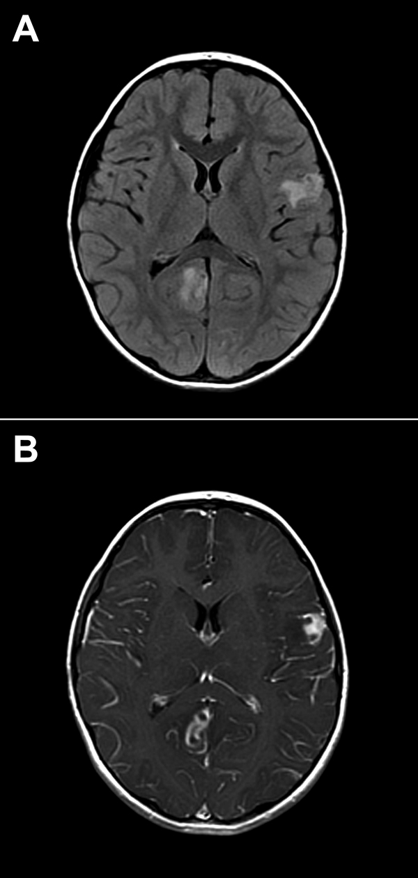
Figure 4. Brain images showing contrast-enhanced lesions in the right occipital and left parietal lobes of a 4-year-old boy with encephalitis caused by infection with Balamuthia mandrillaris amebae. A) T2-weighted fluid-attenuated inversion recovery (FLAIR) image. B) T1-weighted contrasted magnetic resonance image.
Page created: August 13, 2014
Page updated: August 13, 2014
Page reviewed: August 13, 2014
The conclusions, findings, and opinions expressed by authors contributing to this journal do not necessarily reflect the official position of the U.S. Department of Health and Human Services, the Public Health Service, the Centers for Disease Control and Prevention, or the authors' affiliated institutions. Use of trade names is for identification only and does not imply endorsement by any of the groups named above.