Volume 21, Number 1—January 2015
Dispatch
Rapid Diagnostic Tests for Identifying Avian Influenza A(H7N9) Virus in Clinical Samples
Abstract
To determine sensitivity of rapid diagnostic tests for detecting influenza A(H7N9) virus, we compared rapid tests with PCR results and tested different types of clinical samples. Usefulness of seasonal influenza rapid tests for A(H7N9) virus infections is limited because of their low sensitivity for detecting virus in upper respiratory tract specimens.
On March 31, 2013, in China, novel avian influenza A(H7N9) virus infection was diagnosed in 3 persons (1). By October 2013, human infection with influenza A(H7N9) virus had reemerged; the number of cases in this second epidemic wave exceeded that of the first wave (before October 2013) (2). As of March 10, 2014, the virus had caused 379 human cases and 135 human deaths during both epidemic waves in China (2). Because the sensitivity of currently available rapid diagnostic tests (RDTs) for detecting virus in clinical specimens from patients with A(H7N9) virus infection remains largely unknown, we evaluated the sensitivity and specificity of 6 such tests available in China for detecting A(H7N9) virus in different types of clinical specimens from infected patients.
Novel avian influenza A(H7N9) virus has become the most prevalent avian influenza virus strain affecting humans in China. Shortly after the March 2013 outbreak, a real-time reverse transcription PCR (rRT-PCR) for detection of A(H7N9) virus was developed by the Chinese National Influenza Center (3). Although rRT-PCR is now considered the standard laboratory-based assay for detecting influenza virus infections, because of its high sensitivity and specificity, it requires high-level laboratory expertise and might not be available in all locations. Thus, the usefulness of RDTs for detecting A(H7N9) virus infection requires assessment. The sensitivity of 6 RDTs has been evaluated in Australia by using a laboratory influenza A(H7N9) virus isolate shared by the Chinese National Influenza Center and the WHO Collaborating Centre for Reference and Research on Influenza in Melbourne, Australia (4). However, the suitability of RDTs for detecting A(H7N9) virus in clinical specimens from patients remains largely unknown. We therefore evaluated the sensitivity and specificity of 6 RDTs (Table 1) available in China for detecting A(H7N9) virus in different types of clinical specimens.
The RDTs varied according to detection mechanism, time to results, storage temperature, and shelf life. Of the 6 RDTs, 3 were designed to detect influenza A and B viruses, 2 influenza A virus only, and 1 specifically H7 virus (test names and manufacturer information provided in Table 1). We followed manufacturers’ instructions and visually read the results. At the time of the study, 5 of the 6 tests had been approved for detection of seasonal influenza viruses in China, and approval was still pending for the Wondfo H7 test for A(H7N9). Since then, the Wondfo H7 test has been approved by the China Food and Drug Administration.
To evaluate detection limits of the RDTs, we propagated vaccine candidate A(H7N9) virus strain A/Anhui/1/2013 in MDCK cells and determined the mean 50% tissue culture infectious dose (TCID50) per milliliter on the basis of at least 3 independent assays. Viruses were standardized to 1 × 10⁷ TCID50/mL and serially diluted 10-fold in phosphate-buffered saline. The detection limit for 3 RDTs was 103 TCID50/mL and for 2 RDTs was 104 TCID50/mL; 1 RDT could not detect A(H7N9) virus. The following 3 RDTs with the highest sensitivity were chosen for further evaluation of A(H7N9) in clinical specimens: Wantai FluA, Wondfo FluA, and Wondfo H7 (Table 1). The specimens tested were throat swab or sputum (including tracheal aspirates) collected from patients with suspected A(H7N9) virus infection since late March 2013, confirmed by rRT-PCR with primers and probes described previously (1,3), and stored at −80°C.
To compare the efficiency of RDTs for detecting A(H7N9) virus and seasonal influenza A viruses, we also used RDTs and rRT-PCR to test seasonal influenza A(H3N2)–positive and A(H1N1)pdm09-positive throat swab samples collected during January–April 2012. rRT-PCR testing for seasonal influenza virus was conducted according to the World Health Organization protocol (5).
In total, 110 throat swab or sputum specimens from 53 A(H7N9)-infected patients and 115 A(H3N2) and 97 A(H1N1)pdm09 throat swab specimens were tested by using the 3 selected RDTs and rRT-PCR; each specimen was prepared and tested by all 4 assays at the same time. As cycle threshold (Ct) values increased, indicating lower levels of influenza virus in the clinical samples, the sensitivity of RDTs decreased significantly (Table 2). Viral load in throat swab specimens from A(H7N9)-infected patients was significantly lower than that from A(H1N1)pdm09- and A(H3N2)-infected patients (Figure 1).
We then further compared the sensitivity of RDTs for detecting virus in A(H7N9) specimens and seasonal influenza virus specimens with the same influenza A matrix gene Ct intervals. We found that for specimens with Ct <25, RDT sensitivity for A(H7N9) specimens and seasonal influenza virus specimens was similar. However, for specimens with Ct >25, RDT sensitivity was significantly lower when A(H7N9) specimens were compared with seasonal influenza virus specimens with the same Ct interval. Overall, RDT sensitivity for detecting A(H7N9) virus was significantly lower than that for detecting A(H1N1)pdm09 or A(H3N2) viruses (p<0.01). Wantai Flu A and Wondfo Flu A detection of A(H1N1)pdm09 and A(H3N2) viruses did not differ significantly (p>0.05). According to the Wondfo H7 subtype colloidal gold kit, 56 (51%) of the 110 A(H7N9) samples were positive and all 212 A(H1N1)pdm09 (n = 97) and A(H3N2) (n = 115) samples were negative (Table 2), demonstrating that this RDT can distinguish between clinical specimens positive for A(H7N9) and seasonal influenza viruses and that its rate of positivity for detecting A(H7N9) viruses is higher than that of the other 2 RDTs tested (Table 2). Ten throat swab samples that were influenza virus negative by rRT-PCR were also negative by the 3 RDTs.
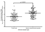
Considering that most A(H7N9) virus–infected patients had pneumonia and that the virus replicates more efficiently in the lower respiratory tract than in the upper respiratory tract (6,7), A(H7N9) viral loads are probably higher in specimens from the lower respiratory tract. Viral loads were significantly higher in sputum/tracheal aspirates than in throat swab samples collected at the same time (Figure 2).
Although most RDTs examined in this study detected not only seasonal influenza virus but also A(H7N9) virus, the sensitivity of RDTs was lower for A(H7N9) virus than for seasonal influenza virus. Even for specimens with the same Ct intervals, RDT sensitivity to A(H7N9) virus was significantly lower than that for either A(H1N1)pdm09 or A(H3N2) virus. The most likely explanation is that cross-reactivity with the nucleocapsid protein–specific antibodies used in RDTs to detect seasonal influenza A virus was significantly lower for A(H7N9) virus. A previous study also indicated that detection sensitivity for swine-origin A(H1N1) viruses varies widely among seasonal influenza A virus RDTs; some tests are unsuitable for detecting several subtypes of avian influenza viruses because of low sensitivity (8).
The Wondfo H7 RDT evaluated in this study was based on a pair of anti-H7 monoclonal antibodies. We found that for each of the 3 Ct intervals, the sensitivity for detecting A(H7N9) was relatively higher for the subtype H7 RDT than for the other RDTs.
Our study indicates that throat swab samples, which have been widely used for influenza diagnosis in China, are not suitable for RDT detection of A(H7N9) virus because of the low levels of virus they contain (Figure 1). Viral loads are significantly higher in sputum samples/tracheal aspirates from the lower respiratory tract than from throat swab samples (Figure 2). If any previously designed influenza A virus–specific RDTs are to be used for detection of A(H7N9) viruses, the kits should be modified for use with sputum and tracheal aspirates by improving extraction. In summary, usefulness of currently available seasonal influenza RDTs for diagnosing A(H7N9) virus infections is limited because of their low sensitivity for detecting virus in upper respiratory tract specimens.
Dr. Y. Chen is a professor at the State Key Laboratory for Diagnosis and Treatment of Infectious Diseases, The First Affiliated Hospital, School of Medicine, Zhejiang University, and belongs to the Collaborative Innovation Center for Diagnosis and Treatment of Infectious Diseases, Hangzhou, Zhejiang, China. His research interests focus on diagnosis and treatment of emerging infectious diseases. Dr. D. Wang is a professor at the National Institute for Viral Disease Control and Prevention, China CDC. Her research focus is influenza surveillance.
Acknowledgments
We thank the National Health and Family Planning Commission of China, Nan-Shan Zhong, and the Chinese Society of Laboratory Medicine.
This study was supported by a special grant for the prevention and control of infectious diseases in China (2012ZX10004-210) and the Key Technologies R&D Program of China (2012BAI37B01).
References
- Gao R, Cao B, Hu Y, Feng Z, Wang D, Hu W, Human infection with a novel avian-origin influenza A (H7N9) virus. N Engl J Med. 2013;368:1888–97 . DOIPubMedGoogle Scholar
- Chinese National Influenza Center. Weekly report, January 2014 [cited 2014 Feb 1]. http://www.cnic.org.cn/uploadfile/2014/0324/20140324095139113.pdf
- World Health Organization. Real-time RT-PCR protocol for the detection of avian influenza A(H7N9) virus [cited 2014 Feb 1]. http://www.who.int/influenza/gisrs_laboratory/cnic_realtime_rt_pcr_protocol_a_h7n9.pdf?ua=1
- Baas C, Barr IG, Fouchier RA, Kelso A, Hurt AC. A comparison of rapid point-of-care tests for the detection of avian influenza A(H7N9) virus, 2013. Euro Surveill. 2013;18:20487 .PubMedGoogle Scholar
- World Health Organization. Manual for the laboratory diagnosis and virological surveillance of influenza [cited 2014 Feb 1]. http://www.who.int/influenza/gisrs_laboratory/manual_diagnosis_surveillance_influenza/en/
- Zhou J, Wang D, Gao R, Zhao B, Song J, Qi X, Biological features of novel avian influenza A (H7N9) virus. Nature. 2013;499:500–3. DOIPubMedGoogle Scholar
- Gao HN, Lu HZ, Cao B, Du B, Shang H, Gan JH, Clinical findings in 111 cases of influenza A (H7N9) virus infection. N Engl J Med. 2013;368:2277–85. DOIPubMedGoogle Scholar
- Sakai-Tagawa Y, Ozawa M, Tamura D, Le QM, Nidom CA, Sugaya N, Sensitivity of influenza rapid diagnostic tests to H5N1 and 2009 pandemic H1N1 viruses. J Clin Microbiol. 2010;48:2872–7 . DOIPubMedGoogle Scholar
Figures
Tables
Cite This Article1These authors contributed equally to this article.
2These authors contributed equally to this article.
Table of Contents – Volume 21, Number 1—January 2015
| EID Search Options |
|---|
|
|
|
|
|
|


Please use the form below to submit correspondence to the authors or contact them at the following address:
Hong Shang, Key Laboratory of AIDS Immunology of Ministry of Health, Department of Laboratory Medicine, The First Hospital of China Medical University, No. 155, Nanjingbei St, Heping District, Shenyang, Liaoning Province 110001, China
Top