Volume 21, Number 10—October 2015
Research
Delayed Disease Progression in Cynomolgus Macaques Infected with Ebola Virus Makona Strain
Andrea Marzi, Friederike Feldmann, Patrick W. Hanley, Dana Scott, Stephan Günther, and Heinz Feldmann
Figure 3
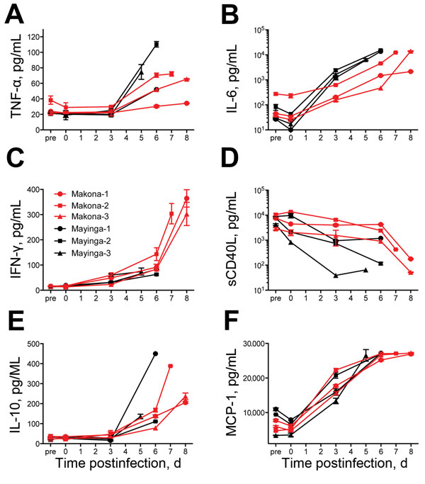
Figure 3. Serum cytokine and chemokine levels for 6 cynomolgus macaques infected with Ebola virus strains Makona or Mayinga. A) Tumor necrosis factor-α (TNF-α); B) Interleukin-6 (IL-6); C) Interferon-γ (IFN-γ); D) Soluble CD40 ligand (sCD40L); E) IL-10; and F) Monocyte chemotactic protein 1 (MCP-1). Kinetics were analyzed in serum samples of each animal collected on days of examination and time of euthanasia.
Page created: September 22, 2015
Page updated: September 22, 2015
Page reviewed: September 22, 2015
The conclusions, findings, and opinions expressed by authors contributing to this journal do not necessarily reflect the official position of the U.S. Department of Health and Human Services, the Public Health Service, the Centers for Disease Control and Prevention, or the authors' affiliated institutions. Use of trade names is for identification only and does not imply endorsement by any of the groups named above.