Volume 21, Number 5—May 2015
CME ACTIVITY - Perspective
Recent US Case of Variant Creutzfeldt-Jakob Disease—Global Implications
Atul Maheshwari , Michael Fischer, Pierluigi Gambetti, Alicia Parker, Aarthi Ram, Claudio Soto, Luis Concha-Marambio, Yvonne Cohen, Ermias D. Belay, Ryan A. Maddox, Simon Mead, Clay Goodman, Joseph S. Kass, Lawrence B. Schonberger, and Haitham M. Hussein
Figure 2
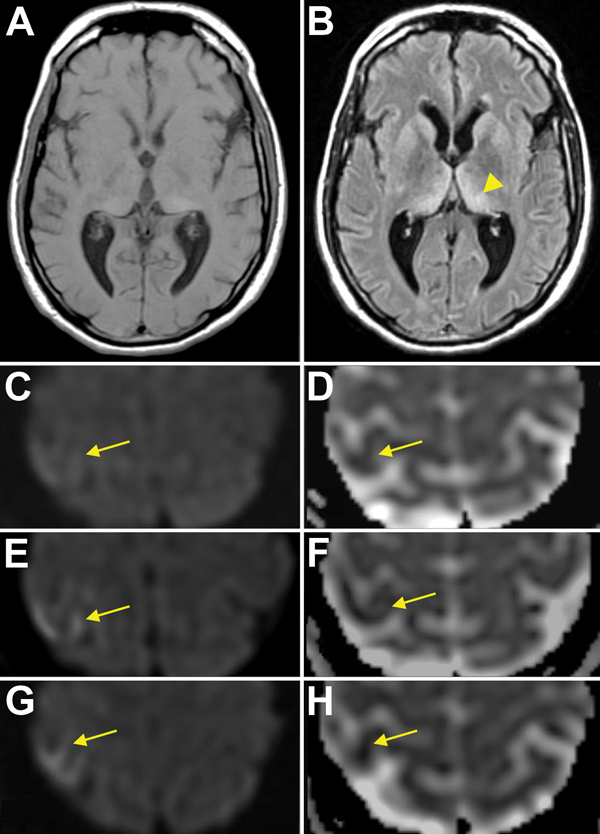
Figure 2. Magnetic resonance imaging (MRI) results for a US patient with variant Creutzfeldt-Jakob disease. T1 sequence (A) and T2 FLAIR sequence (B) show the “pulvinar” or “hockey stick” sign (arrowhead). Initial diffusion weighted imaging (DWI) (C) and apparent diffusion coefficient (ADC) (D) images show subtle restricted diffusion in the right primary motor cortex (arrows). Subsequent MRIs show increasing hyperintensity (arrows) on DWI (E, then G) and further attenuation (arrows) on ADC (F, then H), consistent with the “cortical ribbon” sign.
Page created: April 19, 2015
Page updated: April 19, 2015
Page reviewed: April 19, 2015
The conclusions, findings, and opinions expressed by authors contributing to this journal do not necessarily reflect the official position of the U.S. Department of Health and Human Services, the Public Health Service, the Centers for Disease Control and Prevention, or the authors' affiliated institutions. Use of trade names is for identification only and does not imply endorsement by any of the groups named above.