Volume 21, Number 5—May 2015
Dispatch
Postmortem Stability of Ebola Virus
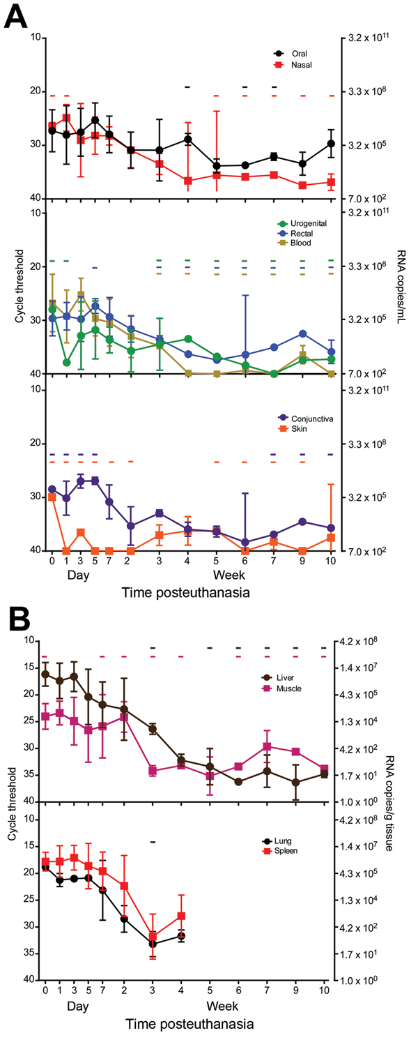
Figure 1

Figure 1. Presence and stability of Ebola virus RNA in deceased cynomolgus macaques. Swab (A) and tissue (B) specimen samples were obtained at the indicated time points, and viral RNA was isolated and used in a 1-step quantitative reverse transcription PCR with a primer/probe set specific for the nucleoprotein gene and standards consisting of known nucleoprotein gene copy numbers. Line plots show means of positive samples from 5 animals up to the 7 day time point and from 3 animals thereafter. Error bars indicate SD, and - indicates time points at which ≥1 animal had undetectable levels of viral RNA. Absence of a hyphen indicates that all animals had detectible levels of viral RNA.
Page created: April 18, 2015
Page updated: April 18, 2015
Page reviewed: April 18, 2015
The conclusions, findings, and opinions expressed by authors contributing to this journal do not necessarily reflect the official position of the U.S. Department of Health and Human Services, the Public Health Service, the Centers for Disease Control and Prevention, or the authors' affiliated institutions. Use of trade names is for identification only and does not imply endorsement by any of the groups named above.