Volume 21, Number 5—May 2015
Research
Novel Thogotovirus Associated with Febrile Illness and Death, United States, 2014
Figure 2

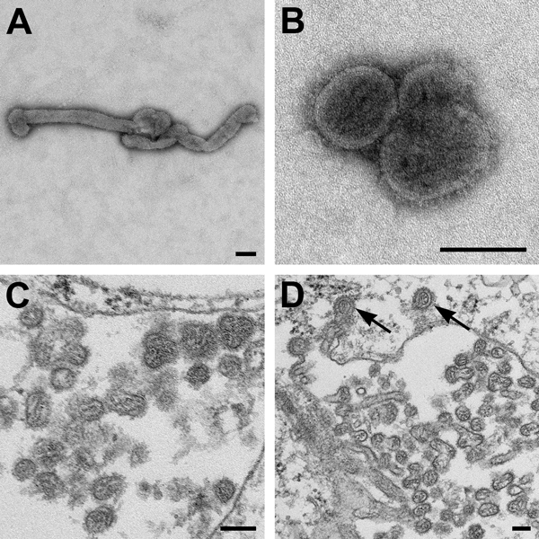
Figure 2. Electron microscopic images of novel Thogotovirus isolate. Filamentous (A) and spherical (B) virus particles with distinct surface projection are visible in culture supernatant that was fixed in 2.5% paraformaldehyde. Thin-section specimens (C and D), fixed in 2.5% glutaraldehyde, show numerous extracellular virions with slices through strands of viral nucleocapsids. Arrows indicate virus particles that have been endocytosed. Scale bars indicate 100 nm.
Page created: April 19, 2015
Page updated: April 19, 2015
Page reviewed: April 19, 2015
The conclusions, findings, and opinions expressed by authors contributing to this journal do not necessarily reflect the official position of the U.S. Department of Health and Human Services, the Public Health Service, the Centers for Disease Control and Prevention, or the authors' affiliated institutions. Use of trade names is for identification only and does not imply endorsement by any of the groups named above.