Skip directly to site content Skip directly to page options Skip directly to A-Z link Skip directly to A-Z link Skip directly to A-Z link
Volume 22, Number 8—August 2016
Synopsis

Co-infections in Visceral Pentastomiasis, Democratic Republic of the Congo

Dennis TappeComments to Author , Mihály Sulyok, Therese Riu, Lajos Rózsa, Imre Bodó, Christoph Schoen, Birgit Muntau, Gergely Babocsay, and Richard Hardi
Author affiliations: Bernhard Nocht Institute, Hamburg, Germany (D. Tappe, B. Muntau); Eberhard Karls University Tübingen, Germany (M. Sulyok); Hôpital Géneral de Reference de Kole, Kole, Democratic Republic of the Congo (T. Riu); MTA-ELTE-MTM Ecology Research Group, Budapest, Hungary (L. Rózsa); Emory University School of Medicine, Atlanta, Georgia, USA (I. Bodó); University of Würzburg, Würzburg, Germany (C. Schoen); Mátra Museum of the Hungarian Natural History Museum, Gyöngyös, Hungary (G. Babocsay); St. Raphael Ophthalmological Center, Mbuji Mayi, Democratic Republic of the Congo (R. Hardi)

Main Article

Figure 2

Histologic and immunohistochemical analyses of resected pentastomid lesions from patients in Sankuru District, Democratic Republic of the Congo, 2014–2015. A) Typical necrotic pentastomid lesion from patient 5. Internal structures of the larvae are decayed; only the directly surrounding exuvia following the annulated body of the parasite and the fibrous capsule are visible. This organism has been moleculary identified as A. armillatus. Periodic acid Schiff stain; original magnification ×2.5. B)

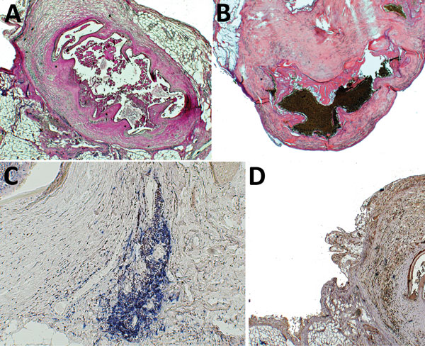
Figure 2. Histologic and immunohistochemical analyses of resected pentastomid lesions from patients in Sankuru District, Democratic Republic of the Congo, 2014–2015. A) Typical necrotic pentastomid lesion from patient 5. Internal structures of the larvae are decayed; only the directly surrounding exuvia following the annulated body of the parasite and the fibrous capsule are visible. This organism has been moleculary identified as A. armillatus. Periodic acid Schiff stain; original magnification ×2.5. B) Necrotic pentastomid lesion from patient 1, with a similar appearance. This organism has been molecularly identified as Raillietiella sp. Hematoxylin and eosin stain; original magnification ×2.5. C) Immunohistochemical staining of B cells (blue) and T cells (brown) surrounding the A. armillatus lesion from patient 5. The lymphocytes cluster locally adjacent to the lesion. Mouse monoclonal anti-CD20 and rabbit monoclonal anti-CD3 stain with hematoxylin counterstain; original magnification ×2.5. D) Immunohistochemical staining for TGF-β (brown) around an A. armillatus larva of patient 5. Immunoreactivity is seen surrounding the lesion and nonspecifically within the parasite larva itself. Rabbit polyclonal IgG with hematoxylin counterstain; original magnification ×2.5.

Main Article

Page created: July 15, 2016
Page updated: July 15, 2016
Page reviewed: July 15, 2016
The conclusions, findings, and opinions expressed by authors contributing to this journal do not necessarily reflect the official position of the U.S. Department of Health and Human Services, the Public Health Service, the Centers for Disease Control and Prevention, or the authors' affiliated institutions. Use of trade names is for identification only and does not imply endorsement by any of the groups named above.
file_external