Volume 23, Number 12—December 2017
Dispatch
Outbreak of Yellow Fever among Nonhuman Primates, Espirito Santo, Brazil, 2017
Figure 2
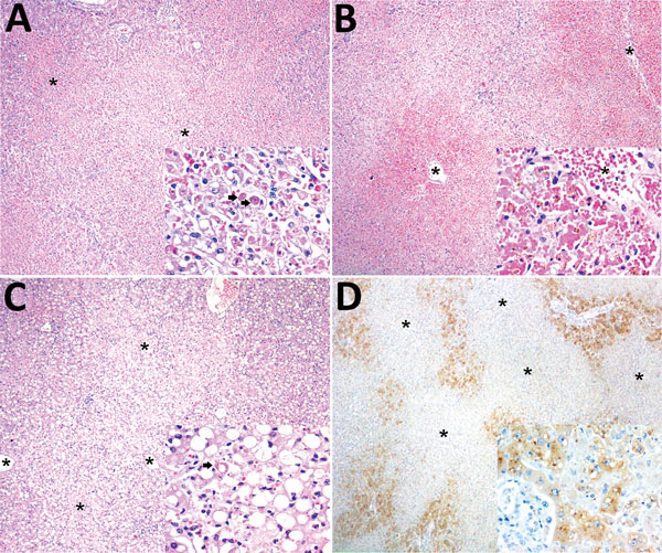
Figure 2. Histopathologic and immunohistochemical findings in the livers of neotropical nonhuman primates that died of yellow fever, Espirito Santo, Brazil, January 2017. Asterisks (*) indicate centrilobular veins. A) Midzonal and centrilobular bridging hepatocellular lytic necrosis. Original magnification ×40; hematoxylin and eosin (H&E) staining. Inset shows lytic hepatocellular necrosis with multiple Councilman-Rocha Lima (apoptotic) bodies (arrows). Original magnification ×400; H&E staining. B) Massive (diffuse) hepatocellular lytic necrosis with severe centrilobular and midzonal hemorrhage. Original magnification ×40; H&E staining. Inset shows prominent hepatocellular necrosis and dropout, and erythrocytes replace the hepatic cords (there is some artifactual formalin pigment [acid hematin] in necrotic hepatocytes). Original magnification ×400; H&E staining. C) Massive macrovacuolar steatosis. Inset shows massive macrovacuolar steatosis mingled with single-cell hepatocellular necrosis (arrow). Original magnification ×400; H&E staining. D) Positive immunolabeling confined to remaining periportal hepatocytes and terminal plate. Original magnification ×40; immunohistochemical staining for yellow fever virus. Inset shows positive granular, cytoplasmic immunolabeling for yellow fever virus antigen in periportal hepatocytes and terminal plate. Original magnification ×400; immunohistochemical staining for yellow fever virus.