Volume 23, Number 3—March 2017
CME ACTIVITY - Synopsis
Three Cases of Neurologic Syndrome Caused by Donor-Derived Microsporidiosis
Rachel Smith , Atis Muehlenbachs, Joanna Schaenmann, Sanjiv Baxi, Sophia Koo, Dianna Blau, Peter Chin-Hong, Anna R. Thorner, Matthew J. Kuehnert, Kristina Wheeler, Alexis Liakos, Jonathan W. Jackson, Theresa Benedict, Alexandre J. da Silva, Jana M. Ritter, Dominique Rollin, Maureen Metcalfe, Cynthia S. Goldsmith, Govinda S. Visvesvara, Sridhar V. Basavaraju, and Sherif Zaki
Figure 3
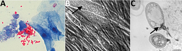
Figure 3. Microsporidia identified in urine samples from liver recipient. A) Urine trichrome stain. Original magnification ×100. B) Cell culture showing microsporidium (arrow). Original magnification ×200. C) Transmission electron microscopic image of infected cell culture with germinating microsporidial spore (arrow). Scale bar indicates 500 nm.
Page created: February 15, 2017
Page updated: February 15, 2017
Page reviewed: February 15, 2017
The conclusions, findings, and opinions expressed by authors contributing to this journal do not necessarily reflect the official position of the U.S. Department of Health and Human Services, the Public Health Service, the Centers for Disease Control and Prevention, or the authors' affiliated institutions. Use of trade names is for identification only and does not imply endorsement by any of the groups named above.