Volume 23, Number 5—May 2017
Dispatch
Virulence Analysis of Bacillus cereus Isolated after Death of Preterm Neonates, Nice, France, 2013
Romain Lotte, Anne-Laure Hérissé, Yasmina Berrouane, Laurène Lotte, Florence Casagrande, Luce Landraud, Sabine Herbin, Nalini Ramarao, Laurent Boyer , and Raymond Ruimy
Figure 1
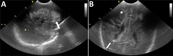
Figure 1. Standard echography cranial ultrasound of premature infant with Bacillus cereus sepsis, Nice, France, 2013. A) Left sagittal section showing large hemorrhagic hyperechogenic area of white material (white arrow). B) Frontal section showing right periventricular kystic hypoechogenic lesions (white arrow) with associated bilateral hemorrhagic hyperechogenic lesions (white star).
Page created: April 17, 2017
Page updated: April 17, 2017
Page reviewed: April 17, 2017
The conclusions, findings, and opinions expressed by authors contributing to this journal do not necessarily reflect the official position of the U.S. Department of Health and Human Services, the Public Health Service, the Centers for Disease Control and Prevention, or the authors' affiliated institutions. Use of trade names is for identification only and does not imply endorsement by any of the groups named above.