Volume 23, Number 6—June 2017
CME ACTIVITY - Synopsis
Sporadic Creutzfeldt-Jakob Disease in 2 Plasma Product Recipients, United Kingdom
Figure
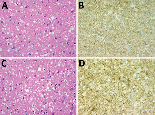
Figure. Results of neuropathologic examinations of the brains of the 2 patients with sporadic Creutzfeldt-Jakob disease, United Kingdom, 2014. A) Microvacuolar spongiform change in the frontal cortex (case 1). Hematoxylin and eosin stain; original magnification ×400. B) Fine granular/synaptic accumulation of abnormal prion protein in the cerebral cortex (case 1). 12F10 antiprion protein antibody; original magnification ×400. C) Microvacuolar spongiform change with neuronal loss and gliosis in the frontal cortex (case 2). Hematoxylin and eosin stain; original magnification ×400. D) Focally intense granular/synaptic accumulation of abnormal prion protein in the cerebral cortex (case 2). 12F10 antiprion protein antibody; original magnification ×400.