Volume 24, Number 5—May 2018
Dispatch
Epizootic Hemorrhagic Disease Virus Serotype 6 Infection in Cattle, Japan, 2015
Yuka Kamomae, Masahiro Kamomae, Yasuyuki Ohta, Mikoto Nabe, Yuichi Kagawa, Yuji Ogura, Tomoko Kato, Shogo Tanaka, Tohru Yanase, and Hiroaki Shirafuji
Figure 1
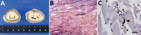
Figure 1. Lesions and epizootic hemorrhagic disease virus serotype 6 (EHDV-6) antigen in esophagus of necropsied cow, Japan, 2015. A) Dilation of lumen. Cross-section of formalin-fixed esophagus of affected cow (right) and control (left). B) Hyaline degeneration of striated muscle accompanied by cell infiltration. Phosphotungstic acid–hematoxylin stain. Scale bar indicates 50 μm. C) EHDV-6 antigen (arrowhead) in vascular endothelium in esophageal muscularis externa. Immunohistochemical stain. Scale bar indicates 20 μm.
Page created: April 17, 2018
Page updated: April 17, 2018
Page reviewed: April 17, 2018
The conclusions, findings, and opinions expressed by authors contributing to this journal do not necessarily reflect the official position of the U.S. Department of Health and Human Services, the Public Health Service, the Centers for Disease Control and Prevention, or the authors' affiliated institutions. Use of trade names is for identification only and does not imply endorsement by any of the groups named above.