Volume 24, Number 6—June 2018
Research
Prion Disease in Dromedary Camels, Algeria
Baaissa Babelhadj, Michele Angelo Di Bari, Laura Pirisinu, Barbara Chiappini, Semir Bechir Suheil Gaouar, Geraldina Riccardi, Stefano Marcon, Umberto Agrimi, Romolo Nonno, and Gabriele Vaccari
Figure 2
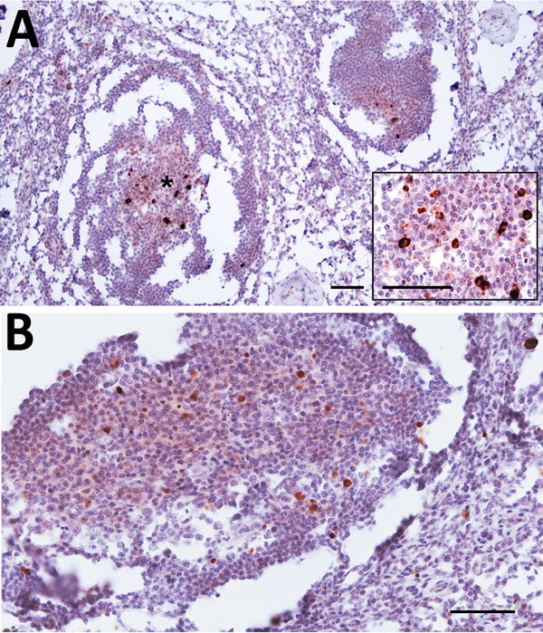
Figure 2. Prion protein immunolabeling in the germinal center of lymphoid follicles of cervical (A) and prescapular (B) lymph nodes of dromedary camel no. 8, Ouargla abattoir, Algeria. The architecture of lymph nodes appears moderately compromised by the partial freezing of samples that accidentally occurred before fixation. Scale bars = 50 μm. Inset in panel A: higher magnification showing the germinal center marked with asterisk; scale bar = 25 mm.
Page created: May 17, 2018
Page updated: May 17, 2018
Page reviewed: May 17, 2018
The conclusions, findings, and opinions expressed by authors contributing to this journal do not necessarily reflect the official position of the U.S. Department of Health and Human Services, the Public Health Service, the Centers for Disease Control and Prevention, or the authors' affiliated institutions. Use of trade names is for identification only and does not imply endorsement by any of the groups named above.