Volume 24, Number 6—June 2018
Research
Prion Disease in Dromedary Camels, Algeria
Figure 3

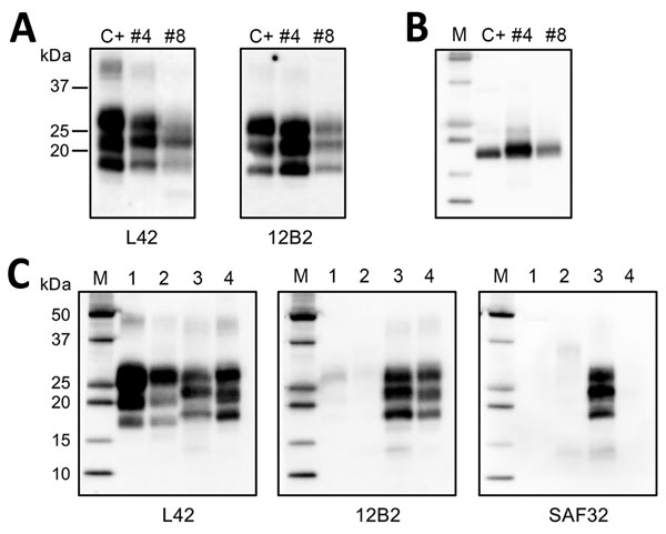
Figure 3. Western blot analysis of protein-resistant core (PrPres) of pathological dromedary prion protein. A) Western blot analysis of proteinase K (PK)–treated PrPSc in brain homogenates from dromedary camels with neurologic symptoms (nos. 4 and 8), Algeria. A sample of sheep scrapie was loaded as control (indicated as C+). Membranes were probed with L42 (left) and 12B2 monoclonal antibody (mAb) (right). Molecular weights (kDa) are indicated on the left. Tissue equivalents loaded per lane were 2 mg for camel samples and 0.1 mg for sheep scrapie. B) Samples after deglycosylation. Membrane was probed with L42 mAb. C) Comparison of dromedary PrPres (from camel no. 4) with sheep bovine spongiform encephalopathy (BSE), bovine BSE, and sheep scrapie samples by ISS (Istituto Superiore di Sanità) discriminatory Western blot (17). Tissue equivalents loaded per lane were 2 mg for dromedary camel and bovine samples and 0.1 mg for sheep samples. In each blot, samples were loaded as follows: lane 1, ovine BSE; lane 2, bovine BSE; lane 3, dromedary camel no. 4; lane 4, ovine scrapie. Membranes were probed with L42, 12B2, and SAF32 mAbs, as indicated. For the analyses in panels B and C, protein standards were loaded and are indicated as M.