Skip directly to site content Skip directly to page options Skip directly to A-Z link Skip directly to A-Z link Skip directly to A-Z link
Volume 24, Number 7—July 2018
Dispatch

Guiana Dolphin Unusual Mortality Event and Link to Cetacean Morbillivirus, Brazil

Kátia R. GrochComments to Author , Elitieri B. Santos-Neto, Josué Díaz-Delgado, Joana M.P. Ikeda, Rafael R. Carvalho, Raissa B. Oliveira, Emi B. Guari, Tatiana L. Bisi, Alexandre F. Azevedo, José Lailson-Brito, and José L. Catão-Dias
Author affiliations: Universidade de São Paulo, São Paulo, Brazil (K.R. Groch, J. Díaz-Delgado, J.L. Catão-Dias); Universidade do Estado do Rio de Janeiro, Rio de Janeiro, Brazil (E.B. Santos-Neto, J.M.P. Ikeda, R.R. Carvalho, R.B. Oliveira, E.B. Guari, T.L. Bisi, A.F. Azevedo, J. Lailson-Brito)

Main Article

Figure 2

Cetacean morbillivirus–associated histopathologic findings in 2 Guiana dolphins (Sotalia guianensis), a female adult (case 1, panels A–C) and a male calf (case 2, panels D–E) A) The mammary gland parenchyma is focally disrupted by lymphohistiocytic inflammatory cells (not visible at this magnification) associated with collapsed and lost acini, and mild fibrosis (asterisks). Original magnification ×40; hematoxylin and eosin (H&E) stain. B) Swollen and degenerating mammary acinar epithelial ce

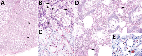
Figure 2. Cetacean morbillivirus–associated histopathologic findings in 2 Guiana dolphins (Sotalia guianensis), a female adult (case 1, panels A–C) and a male calf (case 2, panels D–E). A) The mammary gland parenchyma is focally disrupted by lymphohistiocytic inflammatory cells (not visible at this magnification) associated with collapsed and lost acini, and mild fibrosis (asterisks). Original magnification ×40; hematoxylin and eosin (H&E) stain. B) Swollen and degenerating mammary acinar epithelial cells have numerous intracytoplasmic and intranuclear inclusion bodies (arrows). Original magnification ×200; H&E stain. C) Degenerating and sloughed mammary acinar epithelial cells have intense granular cytoplasmic and intranuclear immunolabeling, identified by immunohistochemistry (IHC) for canine distemper virus (CDV), known to cross react with cetacean morbilliviruses. D) Pulmonary area displaying interstitial pneumonia with mildly thickened alveolar septa and alveoli containing proteinaceous edema, scattered fibrin strands, and small numbers of pleocellular inflammatory cells including occasional syncytia (arrows). Original magnification ×100; H&E stain. E) Degenerating and necrotic type I pneumocytes, sloughed and adhered type II pneumocytes, alveolar and septal macrophages, syncytia (arrow) and circulating (intravascular) mononuclear cells display intense immunolabeling. Original magnification ×400; IHC for CDV.

Main Article

Page created: June 18, 2018
Page updated: June 18, 2018
Page reviewed: June 18, 2018
The conclusions, findings, and opinions expressed by authors contributing to this journal do not necessarily reflect the official position of the U.S. Department of Health and Human Services, the Public Health Service, the Centers for Disease Control and Prevention, or the authors' affiliated institutions. Use of trade names is for identification only and does not imply endorsement by any of the groups named above.
file_external