Volume 24, Number 9—September 2018
Research
Novel Orthopoxvirus and Lethal Disease in Cat, Italy
Figure 2

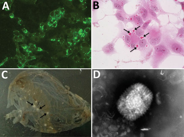
Figure 2. Analysis of an orthopoxvirus isolated from an infected cat, Italy. A) Cytoplasmic fluorescence in infected Vero cells using serum from the diseased cat (original magnification ×400). B) Cytoplasmic inclusion bodies (arrows) in infected Vero cells (hematoxylin and eosin stain, original magnification ×400). C) Pocks (arrows) in the inoculated chorioallantoic membrane of a 12-day-old chick embryo. D) Electron micrograph of orthopoxvirus-like particle from infected Vero cells. The virus preparation was negative-stained with sodium phosphotungstate (original magnification ×25,000).
Page created: August 14, 2018
Page updated: August 14, 2018
Page reviewed: August 14, 2018
The conclusions, findings, and opinions expressed by authors contributing to this journal do not necessarily reflect the official position of the U.S. Department of Health and Human Services, the Public Health Service, the Centers for Disease Control and Prevention, or the authors' affiliated institutions. Use of trade names is for identification only and does not imply endorsement by any of the groups named above.