Volume 24, Number 9—September 2018
Dispatch
Association of Batai Virus Infection and Encephalitis in Harbor Seals, Germany, 2016
Figure 1
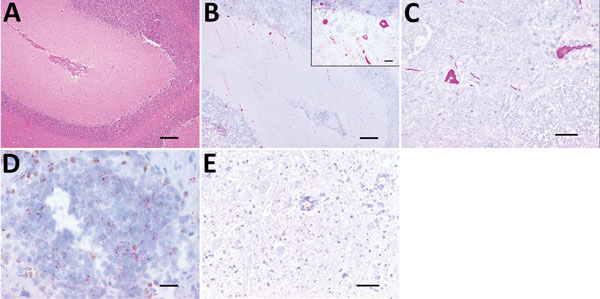
Figure 1. Histologic analysis and fluorescent in situ hybridization (FISH) of a Batai virus BATV)–infected harbor seal, Germany, 2016. A) Cerebellum showing mild to moderate, perivascularly accentuated, lymphohistiocytic inflammation (hematoxylin and eosin stain; scale bar indicates 200 μm). B) Purkinje cells and neurons of granular cell layer showing intracytoplasmic BATV–specific pink, positive result detected by FISH (fast red stain; scale bar indicates 200 µm). Inset: Higher magnification view of analysis using the QuantiGene ViewRNA ISH Tissue 1-Plex Assay Kit and the QuantiGene ViewRNA Chromogenic Signal Amplification Kit (Affymetrix-Panomics, Santa Clara, CA, USA) (fast red stain; scale bar indicates 20 µm). C) Scattered neurons of spinal cord showing a strong, pink, intracytoplasmic BATV-specific result detected by FISH (fast red stain; scale bar indicates 100 µm). D) Cortical and medullary lymphocytes of pulmonary lymph node showing a mild, pink, intracytoplasmic BATV-specific result detected by FISH (fast red stain; scale bar indicates 20 µm). E). Negative control (incubation without probe) of spinal cord showing no BATV-specific result (fast red stain; scale bar indicates 100 µm).
1These authors contributed equally to this article.
2Current affiliation: Utrecht University, Utrecht, the Netherlands.