Volume 25, Number 12—December 2019
Research
Human Infection with Orf Virus and Description of Its Whole Genome, France, 2017
Julien Andreani, Jessica Fongue, Jacques Y. Bou Khalil, Laurene David, Saïd Mougari, Marion Le Bideau, Jonatas Abrahão, Philippe Berbis, and Bernard La Scola
Figure 3
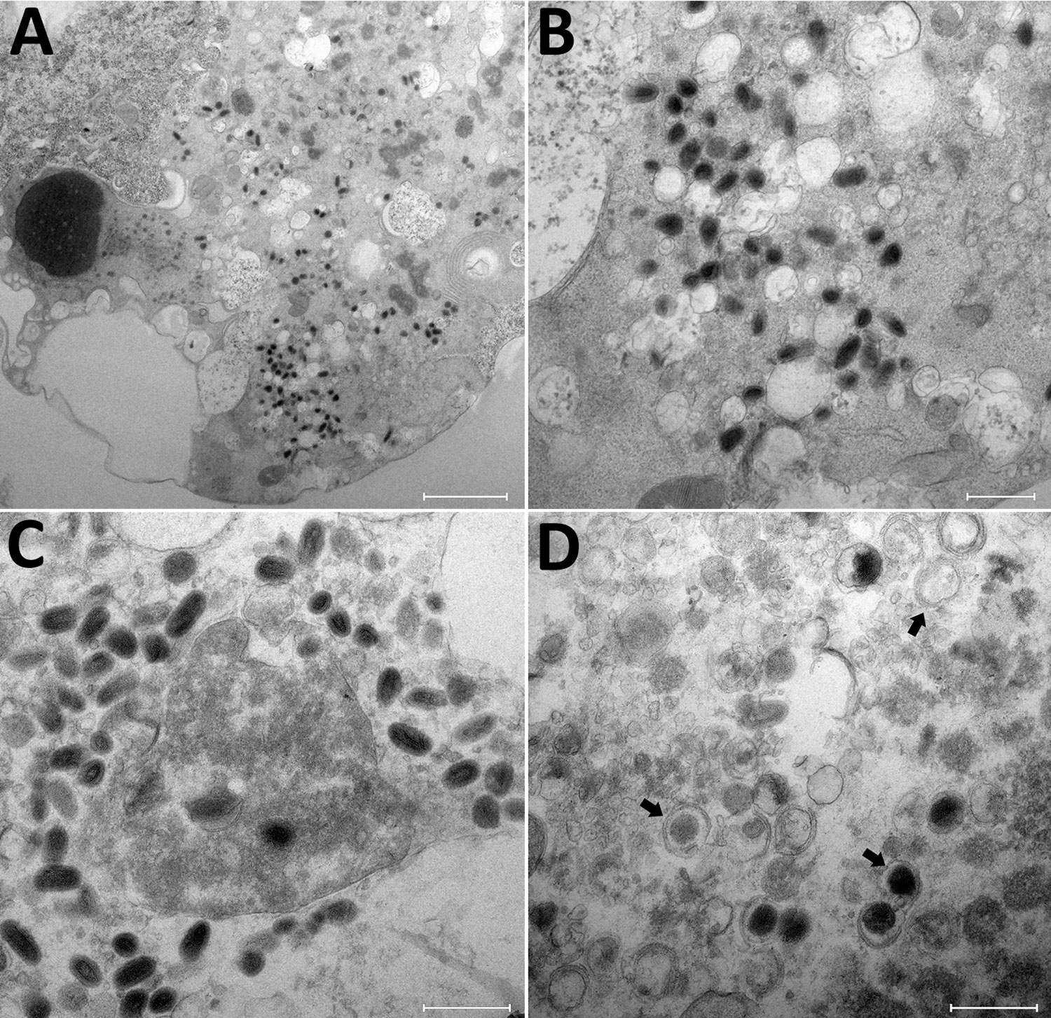
Figure 3. Transmission electron microscopy of OAT3.T cells infected with orf virus IHUMI-1 from a 65-year-old woman in France. A) Ultrathin section of an OAT3.Ts cell at 24 h postinfection harboring orf virus strain IHUMI-1 undergoing its replicative cycle where dense inclusion bodies could be clearly seen in the cell cytoplasm. B, C) Higher magnifications of infected cells showing typical enveloped virions. D) Ultrathin sections of an OAT3.Ts cell showing enveloped particles (arrows). Scale bars indicate 2 μm in panel A, 50 nm in panels B, C, and D.
Page created: November 18, 2019
Page updated: November 18, 2019
Page reviewed: November 18, 2019
The conclusions, findings, and opinions expressed by authors contributing to this journal do not necessarily reflect the official position of the U.S. Department of Health and Human Services, the Public Health Service, the Centers for Disease Control and Prevention, or the authors' affiliated institutions. Use of trade names is for identification only and does not imply endorsement by any of the groups named above.