Volume 26, Number 1—January 2020
Research
High Pathogenicity of Nipah Virus from Pteropus lylei Fruit Bats, Cambodia
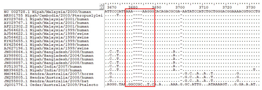
Figure 2

Figure 2. Multiple alignment of the phosphoprotein gene of Nipah virus CSUR381, Cambodia, 2003, and other henipavirus isolates. The highly conserved editing site (5′-AAAAAGGG-3′, red outline) is present in all Nipah and Hendra virus sequences but absent in the nonpathogenic Cedar virus sequence. GenBank accession numbers are provided for all isolates.
Page created: December 18, 2019
Page updated: December 18, 2019
Page reviewed: December 18, 2019
The conclusions, findings, and opinions expressed by authors contributing to this journal do not necessarily reflect the official position of the U.S. Department of Health and Human Services, the Public Health Service, the Centers for Disease Control and Prevention, or the authors' affiliated institutions. Use of trade names is for identification only and does not imply endorsement by any of the groups named above.