Volume 26, Number 7—July 2020
Dispatch
Shuni Virus in Wildlife and Nonequine Domestic Animals, South Africa
Figure 1

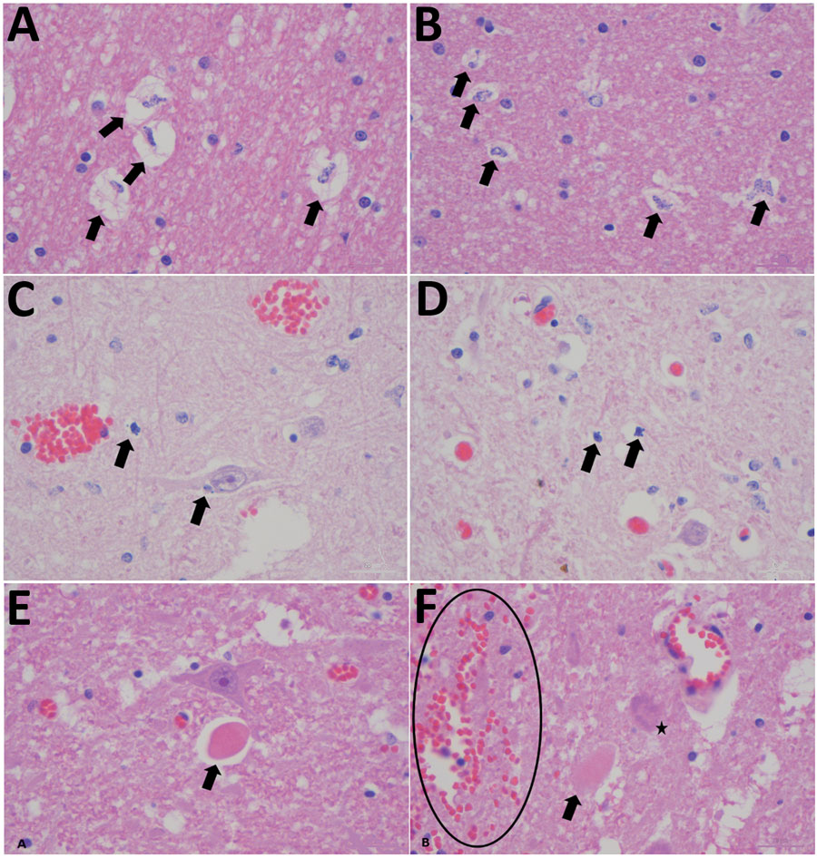
Figure 1. Histopathological changes in formalin-fixed brain tissue of a Shuni virus PCR-positive buffalo (MVA73/10) in South Africa that showed neurologic signs (original magnification 1000×). A, B) Cerebral white matter micro/astrogliosis and cytogenic edema (arrows). C, D) Glial (suspected oligodendroglia) apoptosis (arrows). E, F) Perineural hypereosinophilic bodies (arrows); perivascular and neuropil hemorrhage (circle); single-cell neuronal degeneration (chromatolysis) (star).
Page created: March 25, 2020
Page updated: June 18, 2020
Page reviewed: June 18, 2020
The conclusions, findings, and opinions expressed by authors contributing to this journal do not necessarily reflect the official position of the U.S. Department of Health and Human Services, the Public Health Service, the Centers for Disease Control and Prevention, or the authors' affiliated institutions. Use of trade names is for identification only and does not imply endorsement by any of the groups named above.