Volume 26, Number 9—September 2020
Dispatch
Clinicopathologic and Immunohistochemical Findings from Autopsy of Patient with COVID-19, Japan
Figure 2
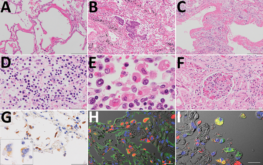
Figure 2. Pathologic findings for the lungs, lymph nodes, and kidneys in an autopsy of an 84-year-old woman who died from coronavirus disease, Toshima Hospital, Tokyo, Japan, February 2020. A) Marked diffuse alveolar damage in exudative phase with prominent hyaline membrane formation in lung tissues. Hematoxylin & eosin (H&E) staining. Scale bar indicates 200 µm. B, C) Desquamation and squamous metaplasia of the epithelium (B) and organized hyaline membranes (C), with septal fibrosis in the organizing phase lesions in lung sections. H&E staining. Scale bar indicates 200 µm. D) Inflammatory infiltrate comprised predominately of plasma cells in the alveolar septa. H&E staining. Scale bar indicates 50 µm. E) Obvious erythrophagocytic macrophages in the lymph nodes. H&E staining. Scale bar indicates 20 µm. F) Numerous microthrombi in the glomerulus in the kidneys. H&E staining. Scale bar indicates 100 µm. G) Immunostaining (brown) of severe acute respiratory syndrome coronavirus 2 antigen in alveolar epithelial cells. Scale bar indicates 50 µm. Inset: multinucleated syncytial cells; scale bar indicates 20 µm. H, I) Double immunofluorescence staining for severe acute respiratory syndrome coronavirus 2 (red) with epithelial cell marker (H; epithelial membrane antigen staining, green); macrophage marker (I; anti-CD68 antibody staining, green) in the same cell. TO-PRO-3 nucleic acid staining (blue) and differential contrast images are also shown. Scale bar indicates 20 µm.