Volume 27, Number 2—February 2021
Dispatch
Plasmodium cynomolgi Co-infections among Symptomatic Malaria Patients, Thailand
Cite This Article
Citation for Media
Abstract
Among 1,180 symptomatic malaria patients, 9 (0.76%) infected with Plasmodium cynomolgi were co-infected with P. vivax (n = 7), P. falciparum (n = 1), or P. vivax and P. knowlesi (n = 1). Patients were from Tak, Chanthaburi, Ubon Ratchathani, Yala, and Narathiwat Provinces, suggesting P. cynomolgi is widespread in this country.
Plasmodium cynomolgi, a simian malaria parasite, possesses biological and genetic characteristics akin to those of the most widespread human malaria parasite, P. vivax. Although P. cynomolgi circulates among monkey species such as long-tailed macaques (Macaca fascicularis) and pig-tailed macaques (M. nemestrina), experimental and accidental transmissions have been implicated in symptomatic infections in humans (1). Several mosquito vectors for human malaria can also transmit P. cynomolgi, posing the risk of cross-species transmission in areas where its natural hosts coexist with people (1,2). Among pig-tailed and long-tailed macaques living in various countries in Southeast Asia, including Thailand, P. cynomolgi infections are not uncommon (3,4). A case of naturally transmitted P. cynomolgi malaria in a human was reported from eastern Malaysia (5). Subsequent surveillance in western Cambodia and northern Sabah state in Malaysia revealed asymptomatic human infection, albeit at low prevalence (6,7). Symptomatic P. cynomolgi infection was diagnosed in a traveler returning to Denmark from Southeast Asia (8). During testing of symptomatic malaria patients in Thailand, we identified 9 co-infected with cryptic P. cynomolgi and other Plasmodium species.
We examined 1,359 blood samples taken from febrile patients who sought treatment at malaria clinics or local hospitals in 5 Thailand provinces: Tak (n = 192, during 2007–2013), Ubon Ratchathani (n = 239, during 2014–2016), Chanthaburi (n = 144, during 2009), Yala (n = 592, during 2008–2018), and Narathiwat (n = 192, during 2008–2010). Using microscopy, we found 1,152 cases in which malaria was caused by P. vivax (869 patients, 75.43%), P. falciparum (272 patients, 23.61%), or co-infection with both species (11 patients, 0.96%). Using species-specific nested PCR, including for P. cynomolgi (Appendix), targeting the mitochondrial cytochrome b gene (mtCytb) of 5 human malaria species for molecular detection, as described elsewhere (9,10), we found malaria in 1,180 patients; P. vivax infections exceeded P. falciparum infections (Table 1). Submicroscopic parasitemia occurred in 28/1,180 (2.4%) patients: 19 infected with P. vivax, 7 with P. falciparum, 1 with P. vivax and P. falciparum, and 1 with P. malariae.
The mean age of all patients was 26.3 (range 7–85) years; 940/1,180 (79.7%) of patients were men. Febrile symptoms, lasting 1–7 days (mean 3.1, SD ±1.3 days) before blood sample collection, developed in all PCR-positive malaria patients. Monoinfection with P. knowlesi occurred in 4 patients, P. malariae in 3, and P. ovale in 1. We detected co-infections in 77 (0.93%) patients; of these co-infections, 55 were P. falciparum and P. vivax. In total (i.e., including both monoinfections and co-infections), P. knowlesi was detected in 18 patients, of which 10 cases were newly identified from Ubon Ratchathani Province, which borders Cambodia and Laos.
We detected P. cynomolgi in 9 patients, all of whom were co-infected with P. vivax (n = 7), P. falciparum (n = 1), or both P. vivax and P. knowlesi (n = 1). The overall prevalence of P. cynomolgi infections was 0.76%. Patients infected with P. cynomolgi were found in all provinces. Although 5 of these patients were from Yala Province, the proportion of P. cynomolgi infections among malaria cases in each malaria-endemic area (0.52%–0.87%) was comparable.
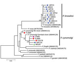
DNA from 10 P. knowlesi isolates from Ubon Ratchathani Province and the 9 P. cynomolgi isolates were subject to nested PCR amplification spanning a 1,318-bp region of mitochondrially encoded cytochrome c oxidase I (mtCOX1). Direct sequencing of the purified PCR-amplified template was successfully performed from all 10 P. knowlesi and from 6 P. cynomolgi isolates. The remaining 3 P. cynomolgi isolates could not be further amplified due to inadequate DNA in the samples. All mtCOX1 sequences of P. knowlesi from Ubon Ratchathani Province were different from one another and distinct from those from the previous case of natural human infection in Thailand (GenBank accession no. AY598141) (11). All 6 amplified P. cynomolgi isolates contained different sequences belonging to 2 clades. One was closely related to the Gombak strain (accession no. AB444129) and the remaining 5 isolates were clustered with the RO strain (accession no. AB444126) (Figure).
All but 1 P. cynomolgi infection occurred in male patients (age 15–53 years, median 32 years). Most P. cynomolgi malaria patients resided in areas where domesticated or wild macaques were living in proximity to humans. Infections with P. cynomolgi occurred in different annual periods; more cases were detected in rainy seasons than in dry seasons (Table 2). The parasite density of P. cynomolgi could not be determined from blood smears because of morphologic resemblance to P. vivax; an isolate co-infected with P. falciparum (YL3634) had very low parasitemia. Of 8 patients with P. cynomolgi co-infection, 6 had parasitemia <10,000 parasites/μL (<0.2% parasitemia). It remains unknown whether P. cynomolgi was co-responsible for symptomatic infections or merely coexisted asymptomatically with other human malaria parasites. However, self-reported defervescence among P. cynomolgi–co-infected patients occurred 1–3 days after antimalarial treatment with chloroquine plus primaquine after onsite microscopic diagnosis of P. vivax malaria or artesunate plus mefloquine for P. falciparum malaria. Unfortunately, data on long-term follow-up were not available.
This report highlights the presence of P. cynomolgi in the human population of Thailand, where natural hosts, both pig-tailed and long-tailed macaques, are prevalent. All patients with P. cynomolgi infections harbored either P. falciparum or P. vivax in their blood, implying that this simian malaria species could share the same anopheline vectors or have different vectors with similar anthropophilic and zoophilic tendencies. The presence of P. cynomolgi in diverse malaria-endemic areas of Thailand suggests that cross-species transmission has occurred. Human infection with P. cynomolgi seems not to be newly emerging because it was detected among blood samples collected over a range of time periods since 2007. Undoubtedly, morphologic similarity between P. cynomolgi and P. vivax can hamper conventional microscopic diagnosis (1,5,8). Cryptic co-existence of simian and human malaria species could further preclude accurate molecular detection when inadequate diagnostic devices are used.
Previous surveys of Plasmodium infections in pig-tailed and long-tailed macaques have revealed the presence of P. cynomolgi and other simian malaria species in Thailand, mainly in the southern part of the country (4). Most patients infected with P. cynomolgi resided in areas where macaques were living in proximity to humans; therefore, the risk of acquiring malaria from this parasite could increase as people encroach into the habitats of infected macaques, as happened with malaria caused by P. knowlesi. Of note, co-infection with P. cynomolgi, P. knowlesi, and P. vivax occurred in a patient in Yala Province whose housing area was surrounded by several domesticated pig-tailed and long-tailed macaques.
Analysis of the mtCOX1 sequences of P. cynomolgi among 6 patients showed that all isolates possessed different genetic sequences, suggesting that several strains or clones of this simian parasite are capable of cross-transmission from macaques to humans. Meanwhile, P. cynomolgi seems to contain 2 divergent lineages (12), represented by RO and Gombak strains. The mtCOX1 sequences of both P. cynomolgi lineages were found in human-derived isolates in this study, further supporting that diverse strains of this parasite can infect people. Likewise, sequence diversity in the mtCOX1 of P. knowlesi from Ubon Ratchathani Province suggests that cross-transmission from macaques to humans may not be restricted to particular parasite strains.
Although human malaria from either parasite may be asymptomatic, infection with P. knowlesi can result in death, but patients infected with P. cynomolgi at worst had only benign symptoms (5–8). However, severe and complicated malaria has been observed in rhesus macaques experimentally infected with P. cynomolgi (13).
Whether severe cynomolgi malaria can occur in humans remains to be elucidated. However, if human infections with P. cynomolgi do become public health problems, diagnostic and control measures might be complicated by the morphological similarity between P. vivax and P. cynomolgi. This possibility makes further surveillance of this simian malaria in humans mandatory.
Dr. Putaporntip is a molecular parasitologist in the Molecular Biology of Malaria and Opportunistic Parasites Research Unit, Department of Parasitology, Faculty of Medicine, Chulalongkorn University, in Bangkok, Thailand. His work focuses on molecular diagnostics, population genetics, and the evolution of malarial parasites in human and nonhuman primates.
Acknowledgments
We are grateful to all patients who provided blood samples and to staff at local malaria clinics and hospitals for assistance in field studies.
Funding for this study was provided to C.P. by the Thailand Research Fund (grant no. RSA5980054), to C.P. by the Asahi Glass Foundation (in fiscal year 2016), and to C.P. and S.J. by the Thai Government Research Budget for Chulalongkorn University (grant nos. GRB-APS-12593011 and GBA-600093004).
References
- Coatney GR, Collins WE, Warren M, Contacos PG. The primate malarias, version 1.0 [CD-ROM] [original book published 1971]. Atlanta: Centers for Disease Control and Prevention; 2003.
- Klein TA, Harrison BA, Dixon SV, Burge JR. Comparative susceptibility of Southeast Asian Anopheles mosquitoes to the simian malaria parasite Plasmodium cynomolgi. J Am Mosq Control Assoc. 1991;7:481–7.PubMedGoogle Scholar
- Putaporntip C, Jongwutiwes S, Thongaree S, Seethamchai S, Grynberg P, Hughes AL. Ecology of malaria parasites infecting Southeast Asian macaques: evidence from cytochrome b sequences. Mol Ecol. 2010;19:3466–76. DOIPubMedGoogle Scholar
- Ta TH, Hisam S, Lanza M, Jiram AI, Ismail N, Rubio JM. First case of a naturally acquired human infection with Plasmodium cynomolgi. Malar J. 2014;13:68. DOIPubMedGoogle Scholar
- Imwong M, Madmanee W, Suwannasin K, Kunasol C, Peto TJ, Tripura R, et al. Asymptomatic natural human infections with the simian malaria parasites Plasmodium cynomolgi and Plasmodium knowlesi. J Infect Dis. 2019;219:695–702. DOIPubMedGoogle Scholar
- Grignard L, Shah S, Chua TH, William T, Drakeley CJ, Fornace KM. Natural human infections with Plasmodium cynomolgi and other malaria species in an elimination setting in Sabah, Malaysia. J Infect Dis. 2019;220:1946–9. DOIPubMedGoogle Scholar
- Hartmeyer GN, Stensvold CR, Fabricius T, Marmolin ES, Hoegh SV, Nielsen HV, et al. Plasmodium cynomolgi as cause of malaria in tourist to Southeast Asia, 2018. Emerg Infect Dis. 2019;25:1936–9. DOIPubMedGoogle Scholar
- Putaporntip C, Buppan P, Jongwutiwes S. Improved performance with saliva and urine as alternative DNA sources for malaria diagnosis by mitochondrial DNA-based PCR assays. Clin Microbiol Infect. 2011;17:1484–91. DOIPubMedGoogle Scholar
- Jongwutiwes S, Buppan P, Kosuvin R, Seethamchai S, Pattanawong U, Sirichaisinthop J, et al. Plasmodium knowlesi Malaria in humans and macaques, Thailand. Emerg Infect Dis. 2011;17:1799–806. DOIPubMedGoogle Scholar
- Jongwutiwes S, Putaporntip C, Iwasaki T, Sata T, Kanbara H. Naturally acquired Plasmodium knowlesi malaria in human, Thailand. Emerg Infect Dis. 2004;10:2211–3. DOIPubMedGoogle Scholar
- Sutton PL, Luo Z, Divis PCS, Friedrich VK, Conway DJ, Singh B, et al. Characterizing the genetic diversity of the monkey malaria parasite Plasmodium cynomolgi. Infect Genet Evol. 2016;40:243–52. DOIPubMedGoogle Scholar
- Joyner CJ, The MaHPIC Consortium, Wood JS, Moreno A, Garcia A, Galinski MR. Case report: severe and complicated cynomolgi malaria in a rhesus macaque resulted in similar histopathological changes as those seen in human malaria. Am J Trop Med Hyg. 2017;97:548–55. DOIPubMedGoogle Scholar
Figure
Tables
Cite This ArticleOriginal Publication Date: January 12, 2021
Table of Contents – Volume 27, Number 2—February 2021
| EID Search Options |
|---|
|
|
|
|
|
|

Please use the form below to submit correspondence to the authors or contact them at the following address:
Chaturong Putaporntip, Molecular Biology of Malaria and Opportunistic Parasites Research Unit, Department of Parasitology, Faculty of Medicine, Chulalongkorn University, Bangkok 10330, Thailand; e-mail:p.chaturong@gmail.com
Top