Volume 27, Number 3—March 2021
Research Letter
Genomic and Pathologic Findings for Prototheca cutis Infection in Cat
Figure
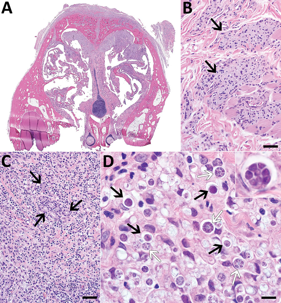
Figure. Histologic lesions associated with protothecosis caused by Prototheca cutis in a cat. A) Subgross cross-section of the nasal turbinates showing marked expansion of the nasal epithelium and overlying subcutaneous tissue. B) Epithelioid macrophages (arrows) with abundant, intracytoplasmic, gray material multifocally dissecting through subdermal collagen and musculature. Hematoxylin and eosin (H&E) stain; scale bar indicates 50 μm. C) Submucosal glands (arrows), markedly displaced by myriad macrophages, neutrophils, lymphocytes, plasma cells, and algal sporangia. H&E stain; scale bar indicates 50 μm. D) Algal sporangia (black arrows), which sometimes endosporulate (white arrows), producing up to 8 endospores (inset). H&E stain; scale bar indicates 10 μm and does not apply to inset.
1These authors contributed equally to this article.