Volume 28, Number 1—January 2022
CME ACTIVITY - Synopsis
Fungal Infections Caused by Kazachstania spp., Strasbourg, France, 2007–2020
Charlotte Kaeuffer, Mathieu Baldacini, Tiffany Ruge, Yvon Ruch, Yves-Jean Zhu, Manon De Cian, Guillaume Philouze, Philippe Bachellier, Julie Denis, Nicolas Lefebvre, Francis Schneider, Yves Hansmann, Valérie Letscher-Bru, Raoul Herbrecht, Marcela Sabou, and François Danion
Figure 2
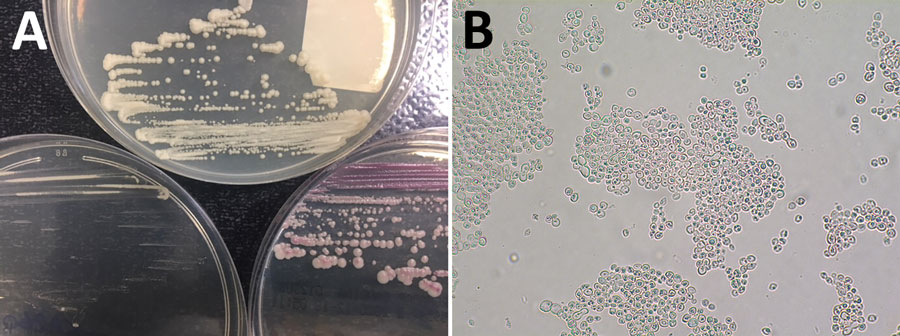
Figure 2. Macroscopic and microscopic examinations of Kazachstania bovina from a patient in Strasbourg, France. A) Macroscopic aspect of K. bovina on 3 agar media: Sabouraud (top), CHROMID Candida (bioMérieux, https://www.biomerieux.fr) (lower left), and CHROMagar Candida (Becton Dickinson, https://www.bd.com) (lower right). B) K. bovina slide-culture on potato carrot bile agar (incubation for 72 h at 27°C, original magnification ×400), showing spherical to ellipsoidal yeast cells with multilateral budding, without filamentation, and some asci containing ascospores.
Page created: November 10, 2021
Page updated: December 17, 2021
Page reviewed: December 17, 2021
The conclusions, findings, and opinions expressed by authors contributing to this journal do not necessarily reflect the official position of the U.S. Department of Health and Human Services, the Public Health Service, the Centers for Disease Control and Prevention, or the authors' affiliated institutions. Use of trade names is for identification only and does not imply endorsement by any of the groups named above.