Volume 28, Number 3—March 2022
Dispatch
Encephalitozoon cuniculi and Extraintestinal Microsporidiosis in Bird Owners
Cite This Article
Citation for Media
Abstract
We identified Encephalitozoon cuniculi genotype II parasites as a cause of extraintestinal microsporidiosis in 2 owners of birds also infected with E. cuniculi. Patients experienced long-lasting nonspecific symptoms; the disease course was more progressive in a patient with diabetes. Our findings suggest direct bird-to-human transmission of this pathogen.
Microsporidia of the genus Encephalitozoon (E. cuniculi, E. hellem, and E. intestinalis) are intracellular pathogens infecting a wide range of animal species. Because spores can be released to the environment via hosts’ feces, urine, and respiratory secretions, they can be ingested or inhaled, posing a risk for zoonotic infection in humans (1). The primary site of Encephalitozoon spp. infection is the small-intestine epithelium, but dissemination and systemic infections are also well known. Infection of a broad spectrum of cell types has been noted, especially for E. cuniculi (1); various pathological changes affecting the digestive, urinary, and respiratory tracts and the nervous system may occur. Because encephalitozoons are opportunistic pathogens, the extraintestinal and disseminated infections and severe symptoms they cause are of concern in immunocompromised hosts, such as transplant recipients or persons living with HIV (2,3). Microsporidiosis develops in patients whose immune response has been weakened by diabetes or malignant disease treated with chemotherapy (2,4).
We describe the case of 2 bird owners in Poland who acquired E. cuniculi–caused microsporidiosis from their infected pet birds. The Human Research Ethics Committee of Wroclaw Medical University (Wroclaw, Poland) approved this study in accordance with agreement no. KB-549/2012. Patients provided written informed consent before examination.
The 2 patients, a woman and a man, both 41 years of age, had nonspecific symptoms of fatigue, exhaustion, joint and muscle pain, frequent colds, and headaches, progressive and more severe in the woman. We observed fever reaching 38°C and lasting several months in both patients. Moreover, the woman had intense night palpitations, symptoms similar to bronchitis (occasionally treated with antimicrobial drugs), blurred vision, dizziness, and impaired concentration. She had had bipolar disorder and diabetes for years. Magnetic resonance imaging scans of her head revealed single minor demyelinating or vascular changes in the white matter of the frontal lobes. No information about diabetes treatment was available. No abnormalities were shown in abdominal ultrasound or chest radiograph. She tested seronegative for Borrelia burgdorferi and Chlamydia psittaci infection. Except for a low leukocyte count (3.00 cells/µL), the woman’s basic laboratory tests of blood and urine and her electrocardiograms showed no abnormalities.
Symptoms emerged in the patients after 2 years of breeding exotic birds together. They had 30 birds of various species: budgerigars, canaries, diamond doves, tricolored parrotfinches, Gouldian finches, and diamond firetails. During the 2-year period, 17 birds died from infectious and metabolic diseases, trauma, and management-related issues.
As a part of standard flock management practice, we performed tentative postmortem diagnostic cytology in 1 tricolored parrotfinch; we suspected disseminated microsporidial infection and conducted a detailed investigation of pooled feces collected from 8 aviaries at the patients’ home and tissues from a dead budgerigar. All samples were delivered on ice to the Laboratory of Veterinary and Medical Protistology, Biology Centre of the Czech Academy of Sciences (České Budějovice, Czech Republic). We collected patients’ urine and stool specimens every 2 days for 1 week, 3 samples from each, during diagnosis and follow-up and examined them at the Department of Biology and Medical Parasitology, Wroclaw Medical University.
We homogenized all samples by mechanical disruption before genomic DNA extraction as described previously (5). We performed nested PCR protocols amplifying a partial sequence of 16S rRNA gene (130 bp), the entire internal transcribed spacer region, and a partial sequence of 5.8S rRNA gene (137–139 bp) of Encephalitozoon spp. (6). In addition, we checked for the presence of Enterocytozoon bieneusi (7) and then performed a phylogenetic analysis of PCR products (5). We used standard light microscopy methods for spore detection (8). We deposited the sequences obtained in this study in GenBank (accession nos. OK356650–60).
We detected microsporidial DNA in the fecal specimens of all birds and in tissue samples from both tested birds. Genotyping revealed the presence of E. hellem genotype 1A in all tested specimens from the lung, liver, duodenum, and jejunum with ileum from the budgerigar, whereas E. cuniculi genotype II was found in pooled feces and in all sections from the liver, gizzard with proventriculus, and duodenum with jejunum and ileum from the tricolored parrotfinch.
Figure
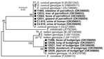
Figure. Phylogenetic relationships of Encephalitozoon cuniculi genotype II and E. hellem genotype 1A obtained from 2 exotic bird breeders and 2 of their birds compared with other ...
E. cuniculi genotype II was present in 2 of the woman’s urine samples and in 1 of the man’s urine samples (Figure). Spores were confirmed in all samples. E. bieneusi was not found. We administered 400 mg albendazole daily for 10 days to both patients. In follow-up examination 3 months after treatment, the patients’ urine repeatedly tested negative for E. cuniculi in all independent samplings, and the patients gradually improved. Patients’ stools remained negative during the entire diagnostic process. No subsequent follow-up was conducted.
Birds are a common source in the propagation of encephalitozoons in the environment (prevalence <15%), either acting as mechanical vectors or developing active infection (9–11). In both wild-living and captive birds, E. hellem is the most prevalent Encephalitozoon species, whereas E. cuniculi has been detected less frequently (9,10). Whether animal hosts indeed propagate microsporidia or serve as transmission vectors is debatable (9). However, the presence of pathogens in both feces and tissues of birds in our study confirms microsporidial proliferation in these animals rather than the passage of spores through the digestive tract; this finding suggests that encephalitozoons may have been circulating in this breeding group for some time. Although most earlier reports demonstrated asymptomatic infections and low infection intensity among birds, intermittent spore shedding in naturally infected birds contaminates the environment (11). Owners could be in constant contact with spores, which highly increases the risk for infection by ingestion or inhalation of spores. As a slow-growing pathogen, E. cuniculi can lead to chronic infection and microsporidiosis.
In immunocompetent hosts, an immune-controlled balance in the host–parasite relationship is established, and extraintestinal infections remain asymptomatic (5). Symptomatic cases of microsporidial infection usually manifest as self-limiting diarrhea (2). Symptomatic extraintestinal Encephalitozoon infections in immunocompetent humans are uncommon and usually present as keratoconjunctivitis (2). Of note, disseminated microsporidiosis caused by E. cuniculi genotype I with involvement of brain and urinary and intestinal tracts has been described in men with type 2 diabetes (4). However, diabetes is considered a risk factor for opportunistic infections. Experimental infection of diabetic mice with E. cuniculi resulted in more symptoms and a higher pathogen burden than in nondiabetic animals (12); indeed, the woman with diabetes in our study experienced more severe symptoms, which led to serious impairments in everyday functioning.
Even though we were able to test only urine and feces and confirmed E. cuniculi in the urinary tract, the symptoms we observed in our patients were complex, indicating disseminated infection. We cannot be confident in the extent to which E. cuniculi infection contributed to these symptoms. However, symptom relief coincided with pathogen clearance after albendazole treatment, which convinced us that microsporidia may have been at least partially involved in symptom development. The severity of symptoms despite the lack of lifelong immunosuppression is puzzling and may arise from high doses of spores acquired as a result of patients’ everyday contact with birds.
In summary, our study documents the risk for bird-to-human transmission of E. cuniculi parasites. Exotic-bird breeders should be aware of the risk for infection with this opportunistic pathogen.
Dr. Kicia is an associate professor in the Department of Biology and Medical Parasitology, Wroclaw Medical University, Wroclaw, Poland. Her research focuses on opportunistic pathogens, including the microsporidia Pneumocystis jirovecii and Cryptosporidium spp. in humans and animals.
Acknowledgment
This work was supported by the Grant Agency of the Czech Republic [grant no. 20-10706S]. The funders had no role in study design, data collection and analysis, decision to publish, or preparation of the manuscript.
References
- Didier ES, Weiss LM. Microsporidiosis: not just in AIDS patients. Curr Opin Infect Dis. 2011;24:490–5. DOIPubMedGoogle Scholar
- Kicia M, Szydłowicz M, Cebulski K, Jakuszko K, Piesiak P, Kowal A, et al. Symptomatic respiratory Encephalitozoon cuniculi infection in renal transplant recipients. Int J Infect Dis. 2019;79:21–5. DOIPubMedGoogle Scholar
- Ditrich O, Chrdle A, Sak B, Chmelík V, Kubále J, Dyková I, et al. Encephalitozoon cuniculi genotype I as a causative agent of brain abscess in an immunocompetent patient. J Clin Microbiol. 2011;49:2769–71. DOIPubMedGoogle Scholar
- Sak B, Kváč M, Kučerová Z, Květoňová D, Saková K. Latent microsporidial infection in immunocompetent individuals - a longitudinal study. PLoS Negl Trop Dis. 2011;5:
e1162 . DOIPubMedGoogle Scholar - Katzwinkel-Wladarsch S, Lieb M, Helse W, Löscher T, Rinder H. Direct amplification and species determination of microsporidian DNA from stool specimens. Trop Med Int Health. 1996;1:373–8. DOIPubMedGoogle Scholar
- Buckholt MA, Lee JH, Tzipori S. Prevalence of Enterocytozoon bieneusi in swine: an 18-month survey at a slaughterhouse in Massachusetts. Appl Environ Microbiol. 2002;68:2595–9. DOIPubMedGoogle Scholar
- Didier ES, Orenstein JM, Aldras A, Bertucci D, Rogers LB, Janney FA. Comparison of three staining methods for detecting microsporidia in fluids. J Clin Microbiol. 1995;33:3138–45. DOIPubMedGoogle Scholar
- Pekmezci D, Yetismis G, Esin C, Duzlu O, Colak ZN, Inci A, et al. Occurrence and molecular identification of zoonotic microsporidia in pet budgerigars (Melopsittacus undulatus) in Turkey. Med Mycol. 2020;59:85–91. DOIPubMedGoogle Scholar
- Hinney B, Sak B, Joachim A, Kváč M. More than a rabbit’s tale - Encephalitozoon spp. in wild mammals and birds. Int J Parasitol Parasites Wildl. 2016;5:76–87. DOIPubMedGoogle Scholar
- Sak B, Kasicková D, Kvác M, Kvetonová D, Ditrich O. Microsporidia in exotic birds: intermittent spore excretion of Encephalitozoon spp. in naturally infected budgerigars (Melopsittacus undulatus). Vet Parasitol. 2010;168:196–200. DOIPubMedGoogle Scholar
- Francisco Neto A, Dell’Armelina Rocha PR, Perez EC, Xavier JG, Peres GB, Spadacci-Morena DD, et al. Diabetes mellitus increases the susceptibility to encephalitozoonosis in mice. PLoS One. 2017;12:
e0186954 . DOIPubMedGoogle Scholar
Figure
Cite This ArticleOriginal Publication Date: February 02, 2022
Table of Contents – Volume 28, Number 3—March 2022
| EID Search Options |
|---|
|
|
|
|
|
|
Please use the form below to submit correspondence to the authors or contact them at the following address:
Marta Kicia, Department of Biology and Medical Parasitology, Wroclaw Medical University, ul. J. Mikulicza-Radeckiego 9, 50-367, Wroclaw, Poland; e-mail:marta.kicia@umw.edu.pl
Top