Skip directly to site content Skip directly to page options Skip directly to A-Z link Skip directly to A-Z link Skip directly to A-Z link
Volume 28, Number 4—April 2022
Research

Fatal Human Alphaherpesvirus 1 Infection in Free-Ranging Black-Tufted Marmosets in Anthropized Environments, Brazil, 2012–2019

Tais M. Wilson, Jana M. Ritter, Roosecelis B. Martines, Hannah A. Bullock, Pamela Fair, Kay W. Radford, Isabel L. Macêdo, Davi E.R. Sousa, Alexandra A.B. Gonçalves, Alessandro P. Romano, Pedro H.O. Passsos, Daniel G. Ramos, Gabriela R.T. Costa, Karina R.L.J. Cavalcante, Cristiano B. de Melo, Sherif R. Zaki, and Marcio B. CastroComments to Author 
Author affiliations: Centers for Disease Control and Prevention, Atlanta, Georgia, USA (T.M. Wilson, J.M. Ritter, R.B. Martines, P. Fair, K.W. Radford, S.R. Zaki); University of Brasília, Brasília, Brazil (T.M. Wilson, I.L. Macêdo, D.E.R. Sousa, A.A.B. Gonçalves, C.B. de Melo, M.B. Castro); Synergy America Inc., Atlanta (H.A. Bullock); Brazilian Ministry of Health, Brasília (A.P. Romano, P.H.O. Passos, D.G. Ramos, K.R.L.J. Cavalcante); Environmental Health Surveillance Directorate of the Federal District, Brasilia (G.R.T. Costa)

Main Article

Figure 3

Pathologic changes in brain of free-ranging black-tufted marmosets with fatal human alphaherpesvirus 1 infection, Brazil, 2012–2019. A) Necrotizing meningoencephalitis. Hemotoxylin and eosin (H&E) stain; original magnification ×10. B) Neuronal degeneration and glial nodule. H&E stain; original magnification ×40. C) Neuronal necrosis with microglial proliferation and expansion of Virchow–Robbin spaces by lymphocytes, histiocytes, and few plasma cells. Neurons and glial cells show intranuclear inclusion bodies and prominent margination of the nuclear chromatin. H&E stain; original magnification ×63. D) Prominent neutrophilic inflammation accompanies neuronal necrosis and intranuclear inclusion bodies. H&E stain; original magnification ×63. E) Human alphaherpesvirus 1 immunostaining within neurons (immunohistochemistry; original magnification ×40). F) Intranuclear (arrowhead) and cytoplasmic (arrow) herpesvirus particles in gray matter. Transmission electron microscopy; scale bar indicate 500 nm. Inset: cytoplasmic herpesvirus particles (arrow) white matter, myelinated axon (arrowhead); scale bar indicates 200 nm.

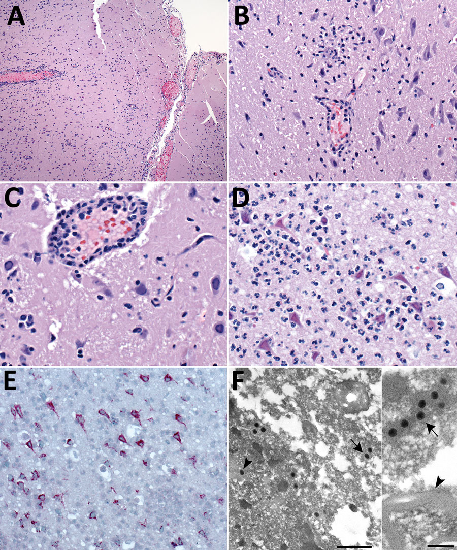
Figure 3. Pathologic changes in brain of free-ranging black-tufted marmosets with fatal human alphaherpesvirus 1 infection, Brazil, 2012–2019. A) Necrotizing meningoencephalitis. Hemotoxylin and eosin (H&E) stain; original magnification ×10. B) Neuronal degeneration and glial nodule. H&E stain; original magnification ×40. C) Neuronal necrosis with microglial proliferation and expansion of Virchow–Robbin spaces by lymphocytes, histiocytes, and few plasma cells. Neurons and glial cells show intranuclear inclusion bodies and prominent margination of the nuclear chromatin. H&E stain; original magnification ×63. D) Prominent neutrophilic inflammation accompanies neuronal necrosis and intranuclear inclusion bodies. H&E stain; original magnification ×63. E) Human alphaherpesvirus 1 immunostaining within neurons (immunohistochemistry; original magnification ×40). F) Intranuclear (arrowhead) and cytoplasmic (arrow) herpesvirus particles in gray matter. Transmission electron microscopy; scale bar indicate 500 nm. Inset: cytoplasmic herpesvirus particles (arrow) white matter, myelinated axon (arrowhead); scale bar indicates 200 nm.

Main Article

Page created: January 26, 2022
Page updated: March 19, 2022
Page reviewed: March 19, 2022
The conclusions, findings, and opinions expressed by authors contributing to this journal do not necessarily reflect the official position of the U.S. Department of Health and Human Services, the Public Health Service, the Centers for Disease Control and Prevention, or the authors' affiliated institutions. Use of trade names is for identification only and does not imply endorsement by any of the groups named above.
file_external