Fatal Human Alphaherpesvirus 1 Infection in Free-Ranging Black-Tufted Marmosets in Anthropized Environments, Brazil, 2012–2019
Tais M. Wilson, Jana M. Ritter, Roosecelis B. Martines, Hannah A. Bullock, Pamela Fair, Kay W. Radford, Isabel L. Macêdo, Davi E.R. Sousa, Alexandra A.B. Gonçalves, Alessandro P. Romano, Pedro H.O. Passsos, Daniel G. Ramos, Gabriela R.T. Costa, Karina R.L.J. Cavalcante, Cristiano B. de Melo, Sherif R. Zaki, and Marcio B. Castro

Author affiliations: Centers for Disease Control and Prevention, Atlanta, Georgia, USA (T.M. Wilson, J.M. Ritter, R.B. Martines, P. Fair, K.W. Radford, S.R. Zaki); University of Brasília, Brasília, Brazil (T.M. Wilson, I.L. Macêdo, D.E.R. Sousa, A.A.B. Gonçalves, C.B. de Melo, M.B. Castro); Synergy America Inc., Atlanta (H.A. Bullock); Brazilian Ministry of Health, Brasília (A.P. Romano, P.H.O. Passos, D.G. Ramos, K.R.L.J. Cavalcante); Environmental Health Surveillance Directorate of the Federal District, Brasilia (G.R.T. Costa)
Main Article
Figure 4
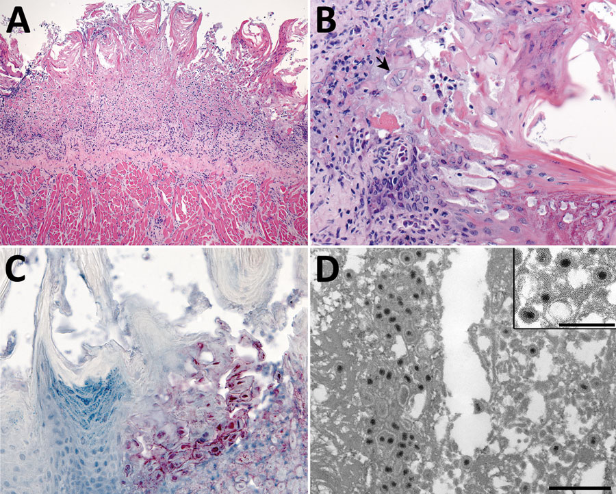
Figure 4. Tongue pathology in free-ranging black-tufted marmosets with fatal human alphaherpesvirus 1 infection, Brazil, 2012–2019. A) Severe necrosis of epithelium. Hemotoxylin and eosin (H&E) stain; original magnification ×10. B) Intranuclear inclusion bodies in epithelial cells at the margin of the lesion and multinucleated syncytial cell (arrow). H&E stain; original magnification ×40. C) Human alphaherpesvirus 1 immunostaining within epithelial cells in the area of necrotizing glossitis. Immunihistochemistry; original magnification ×40. D) Epithelial cell containing accumulations of herpesvirus within the cytoplasm. Transmission electron microscopy; scale bar indicates 600 nm. Inset: higher magnification image of herpesvirus particles with well-defined tegument layer in the cytoplasm; scale bar indicates 400 nm.
Main Article
Page created: January 26, 2022
Page updated: March 19, 2022
Page reviewed: March 19, 2022
The conclusions, findings, and opinions expressed by authors contributing to this journal do not necessarily reflect the official position of the U.S. Department of Health and Human Services, the Public Health Service, the Centers for Disease Control and Prevention, or the authors' affiliated institutions. Use of trade names is for identification only and does not imply endorsement by any of the groups named above.