Volume 28, Number 7—July 2022
Dispatch
Determining Infected Aortic Aneurysm Treatment Using Focused Detection of Helicobacter cinaedi
Saito Jien , Rimbara Emiko, Inaguma Shingo, Hasegawa Chihiro, Kamiya Shinji, Mizuno Akihiro, Sone Yoshiaki, Ogawa Tatsuhito, Numata Yukihide, Takahashi Satoru, and Asano Miki
Figure 1
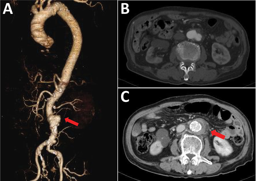
Figure 1. Contrast-enhanced computed tomography imaging for Case-patient 3 in the Helicobacter cinaedi group of 10 patients with infected aortic aneurysms with or without H. cinaedi, Aichi, Japan, September 2017–January 2021. A, B) The infrarenal aortic aneurysm had a maximum short diameter of 39 mm and a cystic protrusion of 19 mm (arrow in panel A) before the operation. C) After the operation, the adipose tissue concentration increased around the aneurysm (arrow).
Page created: May 05, 2022
Page updated: June 18, 2022
Page reviewed: June 18, 2022
The conclusions, findings, and opinions expressed by authors contributing to this journal do not necessarily reflect the official position of the U.S. Department of Health and Human Services, the Public Health Service, the Centers for Disease Control and Prevention, or the authors' affiliated institutions. Use of trade names is for identification only and does not imply endorsement by any of the groups named above.