Volume 28, Number 7—July 2022
Dispatch
Determining Infected Aortic Aneurysm Treatment Using Focused Detection of Helicobacter cinaedi
Figure 2
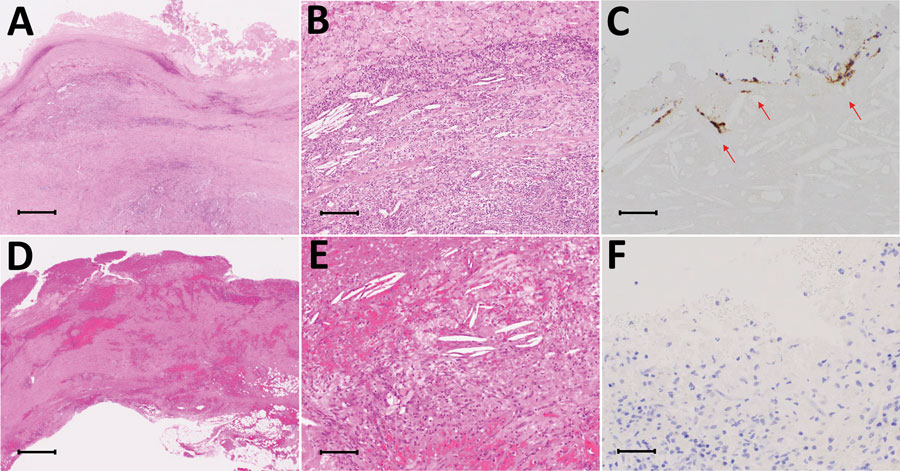
Figure 2. Comparison of images from patients in the Helicobacter cinaedi group with patients from the non–H. cinaedi group among 10 patients with infected aortic aneurysms with or without H. cinaedi, Aichi, Japan, September 2017–January 2021. Immunohistochemistry was performed on the whole cell lysates of H. cinaedi strain MRY08-1234 isolated from immunocompromised patients in Japan by raising anti–rabbit H. cinaedi IgG. One of 2 case-patients with resected tissue in the H. cinaedi group had positive immunostaining (patient 1). A–C) Case-patient 1 in the H. cinaedi group. D–F) Case-patient 4 in the non–H. cinaedi group. In images from both patients, lymphocyte and neutrophil infiltrates, cholesterol clefts, foam cells, plasma cells, foreign body giant cells, and hemosiderin deposition are visible (A, B, D, E; hematoxylin & eosin). Immunohistochemistry stain shows of H. cinaedi organisms in the aortic intima (arrow in C) and negative results (F). Scale bars: 1,000 µm in A, D; 100 µm in B, E; 50 µm in C, F.