Volume 28, Number 9—September 2022
Research Letter
Trichodysplasia Spinulosa Polyomavirus Endothelial Infection, California, USA
Figure

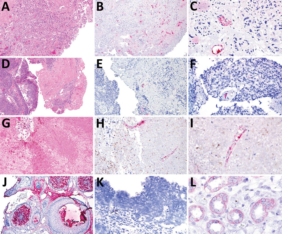
Figure. Tissue samples from 3 patients with trichodysplasia spinulosa polyomavirus endothelial infection, California, USA. We performed hematoxylin and eosin (H&E) staining and RNAScope in situ hybridization (ISH) to detect trichodysplasia spinulosa polyomavirus (TSPyV) in formalin-fixed, paraffin-embedded tissue specimens. Bright red, granular staining in endothelium indicates TSPyV RNA. A) Case 1, H&E staining, original magnification ×10; B) case 1, TSPyV ISH, original magnification ×10; C) case 1, TSPyV ISH, original magnification ×40; D) case 2, H&E staining, original magnification ×5; E) case 2, TSPyV ISH, original magnification ×10; F) case 2, TSPyV ISH, original magnification ×40; G) case 3, H&E staining, original magnification ×5; H) case 3, TSPyV ISH, original magnification ×10; I) case 3, TSPyV ISH, original magnification ×40; J) biopsy from patient with cutaneous trichodysplasia spinulosa (positive control), TSPyV ISH, original magnification ×10; K) cutaneous biopsy from patient with Merkel cell polyomavirus-positive Merkel cell carcinoma (negative control), TSPyV ISH, original magnification ×40; L) renal biopsy from patient with BK polyomavirus nephropathy (negative control), TSPyV ISH, original magnification ×60.
1Current affiliation: Guardant Health, Redwood City, California, USA.