Volume 29, Number 1—January 2023
Dispatch
Pathologic and Immunohistochemical Evidence of Possible Francisellaceae among Aborted Ovine Fetuses, Uruguay
Federico Giannitti , Matías A. Dorsch, Carlos O. Schild, Rubén D. Caffarena, Karen Sverlow, Aníbal G. Armién, and Franklin Riet-Correa
Figure 1
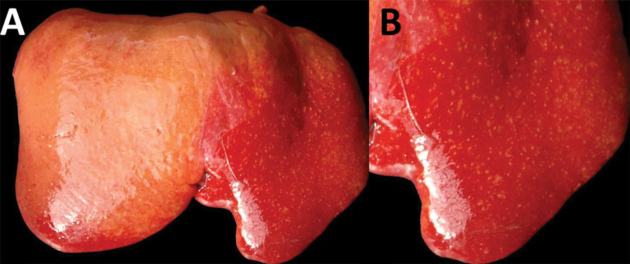
Figure 1. Diaphragmatic view of diseased liver from an aborted ovine fetus (fetus B) with possible Francisellaceae infection, Uruguay. A) Myriad discrete, white to yellowish, round, coalescing foci are visible, ranging from pinpoint to ≈2 mm in diameter, with a multifocal disseminated distribution in the hepatic parenchyma indicative of necrotizing hepatitis. Lesions are more visible in the left liver lobe (right side of the image) than the right liver lobe (left side of the image), in which the hepatic parenchyma is diffusely pale due to moderate autolysis. B) Isolated view of left liver lobe with disseminated foci of necrotizing hepatitis, which are characteristic in cases of tularemia.
Page created: November 04, 2022
Page updated: December 22, 2022
Page reviewed: December 22, 2022
The conclusions, findings, and opinions expressed by authors contributing to this journal do not necessarily reflect the official position of the U.S. Department of Health and Human Services, the Public Health Service, the Centers for Disease Control and Prevention, or the authors' affiliated institutions. Use of trade names is for identification only and does not imply endorsement by any of the groups named above.