Volume 29, Number 1—January 2023
Dispatch
Pathologic and Immunohistochemical Evidence of Possible Francisellaceae among Aborted Ovine Fetuses, Uruguay
Figure 2
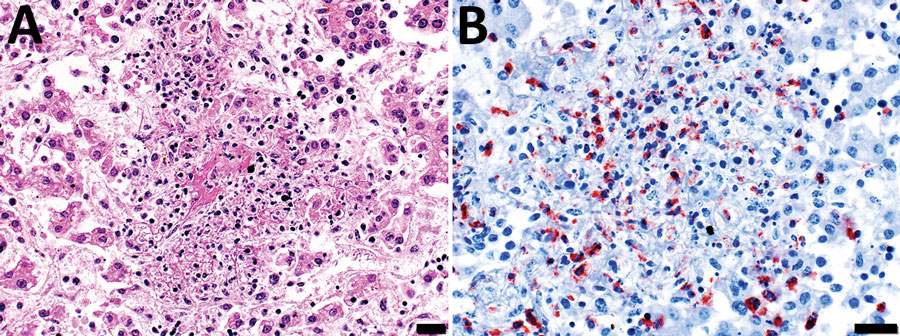
Figure 2. Microscopic images of diseased liver from an aborted ovine (fetus B) with possible Francisellaceae infection, Uruguay. A) Hematoxylin and eosin stain of section of liver. Hepatic histoarchitecture in center of image is effaced by eosinophilic karyorrhectic cellular debris (i.e., necrosis), fibrin exudate, and inflammatory cell infiltrates, mostly neutrophils and macrophages, evidence of severe necrotizing fibrinosuppurative hepatitis. Scale bar indicates 20 μm. B) Immunohistochemistry using a mouse monoclonal antibody raised against F. tularensis lipopolysaccharide and hematoxylin counterstain of section of liver shows abundant intralesional intracellular and extracellular immunoreactivity as a granular brownish-red precipitate (chromogen 3-amino-9-ethylcarbazole). Intracytoplasmic immunoreactivity was noted in infiltrating neutrophils and macrophages. Scale bar indicates 20 μm.