Volume 29, Number 11—November 2023
Research
Neurologic Effects of SARS-CoV-2 Transmitted among Dogs
Figure 3
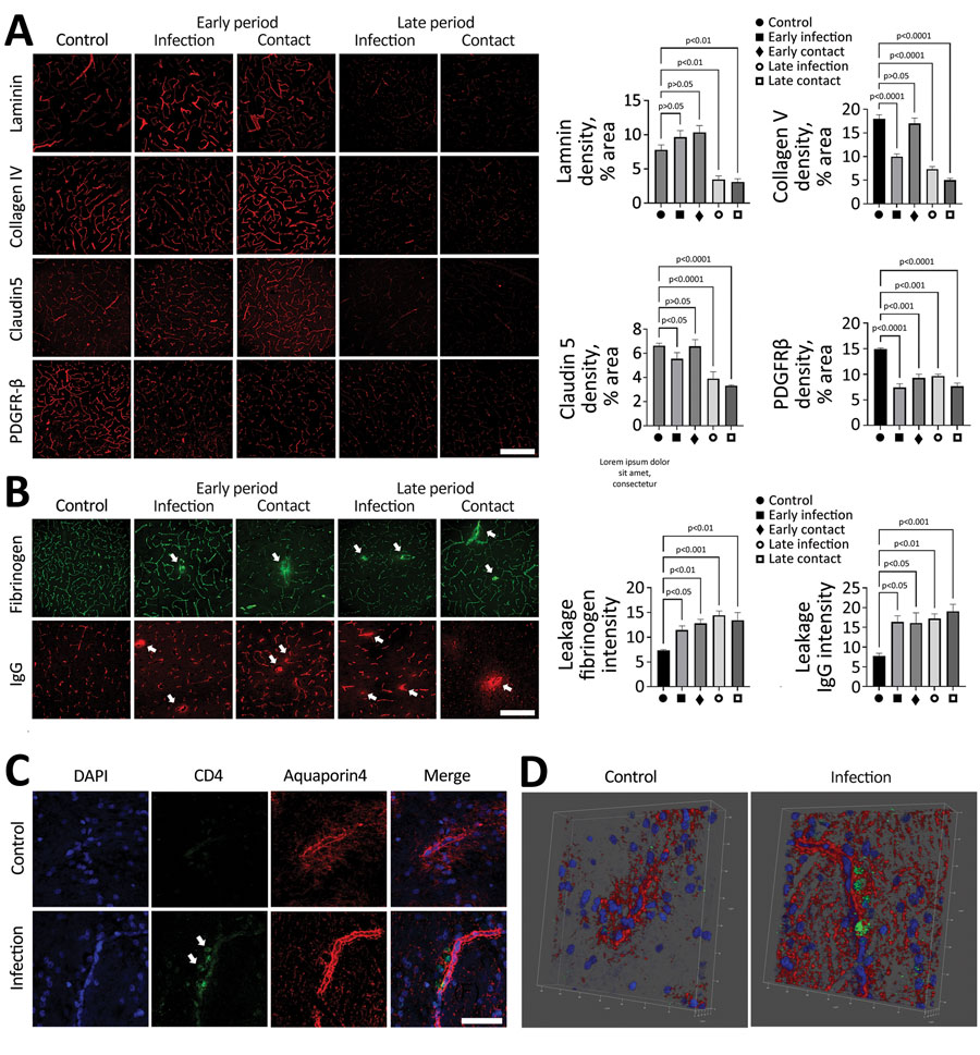
Figure 3. SARS-CoV-2 infection disrupting the blood–brain barrier (BBB) and immune cells infiltrating the brains of infected dogs in study of the neurologic effects of SARS-CoV-2 transmitted among dogs. A) Representative fluorescent images and statistical results of vascular markers including laminin, claudin 5, collagen IV, and PDGFR-β staining of canine brain sections derived from SARS-CoV-2–infected and contact dogs at early and late days after infection. Scale bar indicates 200 μm. B) Brain sections from SARS-CoV-2–infected and contact dogs stained with fibrinogen (green) and IgG (red), which represent BBB leakage staining, at early and late days after infection. Scale bar indicates 200 μm. (C) Representative fluorescent images of the brain sections stained with the CD4 (green) and aquaporin 4 (red), which are markers of CD4-positive T cells and astrocytic end foot, respectively. The CD4-positive cells were infiltrated in the brain parenchyma (white arrows) in the SARS-CoV-2–infected group. Scale bar indicates 50 μm. (D) Representative 3D images of CD4 (green) and aquaporin 4 (red). Statistical significance was determined using a 1-way analysis of variance. Data are presented as mean ±SEM.
1These authors contributed equally to this article.