Volume 29, Number 11—November 2023
Research
Neurologic Effects of SARS-CoV-2 Transmitted among Dogs
Figure 4
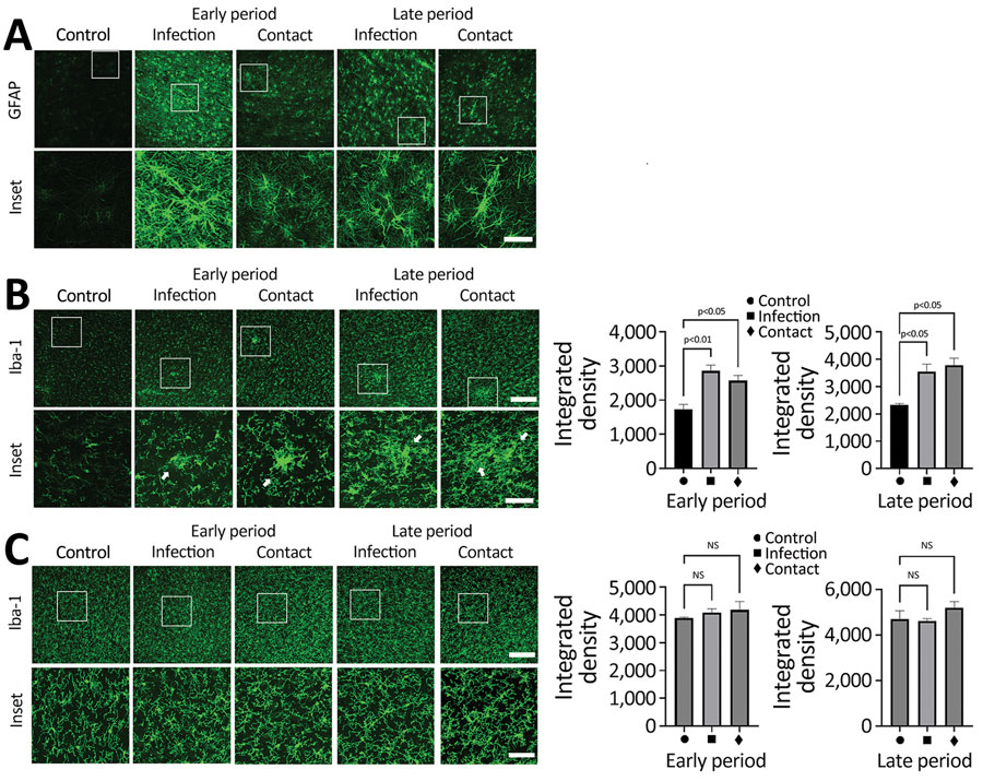
Figure 4. SARS-CoV-2 induces activation of microglial cells in the brain white matter in a region-specific manner in SARS-CoV-2–infected and contact dogs in study of the neurologic effects of SARS-CoV-2 transmitted among dogs. A) Representative fluorescent images of glial fibrillary acidic protein (activation astrocyte marker, green) staining of canine brain sections derived from SARS-CoV-2–infected and contact groups at early and late days after infection. Scale bars indicate 200 μm; in insets, 50 μm. B) Representative fluorescent images and statistical results of Iba-1 (a marker of microglia; green) staining of canine brain white matter sections derived from SARS-CoV-2–infected and contact dogs at early and late dpi. Scale bars indicate 200 μm; in insets, 50 μm. C) Representative fluorescent images and statistical results of Iba-1 (a marker of microglia, green) staining of canine brain gray matter sections derived from SARS-CoV-2–infected and contact dogs at early and late dpi. Scale bars indicate 200 μm; in insets, 50 μm. Statistical significance was determined using a 1-way analysis of variance. Data in graphs are presented as means ±SEM.
1These authors contributed equally to this article.