Volume 29, Number 12—December 2023
Research
Fatal Human Neurologic Infection Caused by Pigeon Avian Paramyxovirus-1, Australia
Siobhan Hurley1 , John Sebastian Eden1, John Bingham, Michael Rodriguez, Matthew J. Neave, Alexandra Johnson, Annaleise R. Howard-Jones, Jen Kok, Antoinette Anazodo, Brendan McMullan, David T. Williams, James Watson, Annalisa Solinas, Ki Wook Kim2, and William Rawlinson2
Figure 2
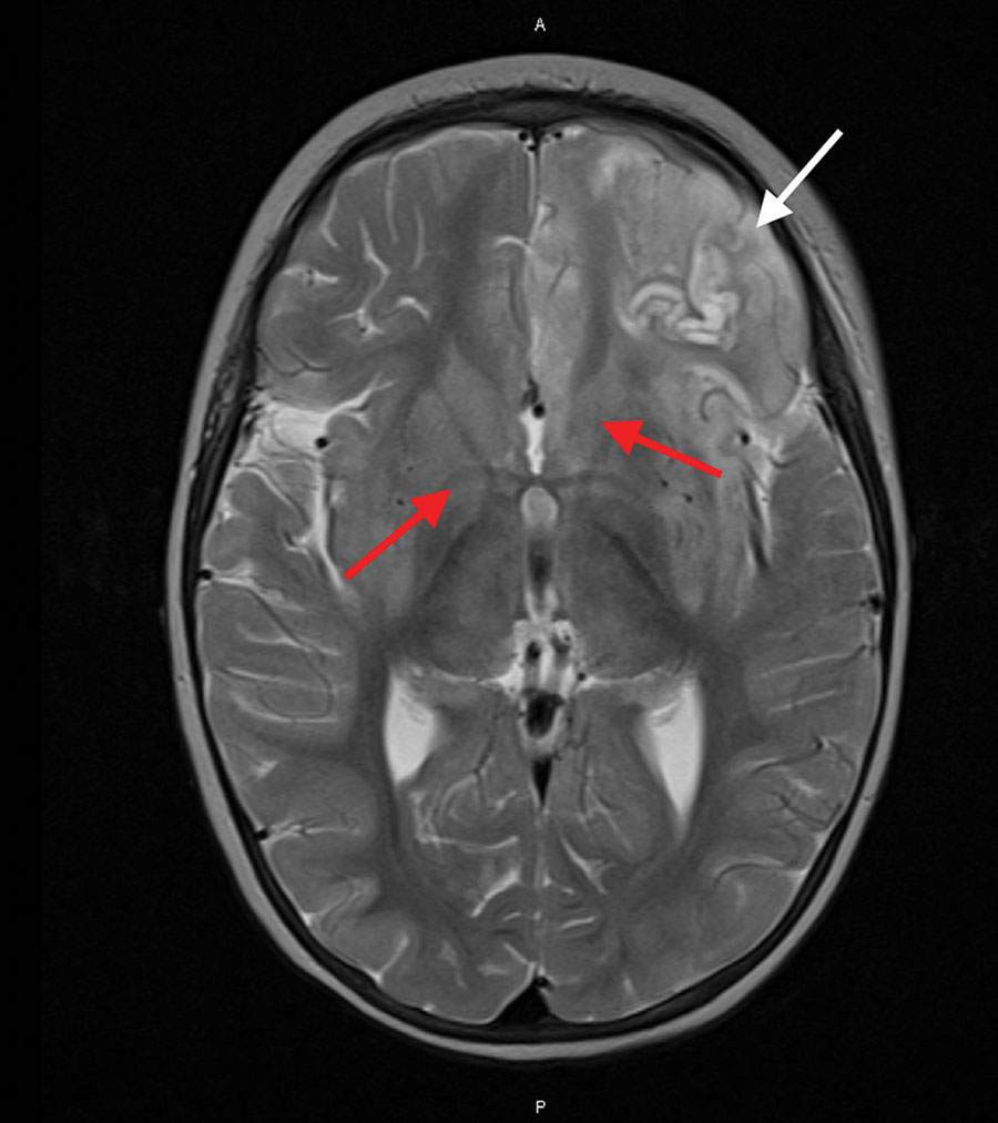
Figure 2. Magnetic resonance imaging of the brain of an immunocompromised child with avian paramyxovirus type 1 infection, Australia. Image, captured 16 days after hospital admission, shows predominantly left frontal and insular T2 signal hyperintensity evolving into laminar necrosis (white arrow) and hyperintensity of deep gray-matter structures (red arrows).
1These first authors contributed equally to this article.
2These senior authors contributed equally to this article.
Page created: November 13, 2023
Page updated: November 18, 2023
Page reviewed: November 18, 2023
The conclusions, findings, and opinions expressed by authors contributing to this journal do not necessarily reflect the official position of the U.S. Department of Health and Human Services, the Public Health Service, the Centers for Disease Control and Prevention, or the authors' affiliated institutions. Use of trade names is for identification only and does not imply endorsement by any of the groups named above.