Volume 29, Number 3—March 2023
Research Letter
Tick-Borne Encephalitis in Pregnant Woman and Long-Term Sequelae
Cite This Article
Citation for Media
Abstract
We report a case of severe tick-borne encephalitis in a pregnant woman, leading to a prolonged stay in the intensive care unit. She showed minor clinical improvement >6 months after her presumed infection. The patient was not vaccinated, although an effective vaccine is available and not contraindicated during pregnancy.
Tick-borne encephalitis (TBE), an emerging infectious disease, has shown a deeply evolving epidemiology during the past decade, especially in Europe (1). TBE virus (TBEV) is transmitted mainly to humans by tick bites and occasionally by consumption of contaminated dairy products (1). Although most infections caused by the TBEV European subtype are asymptomatic, some patients’ conditions could worsen to show severe encephalitis, associated with long-term sequelae (1). Data dealing with TBEV infection during pregnancy are scarce. We report a case of severe TBE and long-term sequelae in a pregnant woman.
In July 2020, a 34-year-old woman at 20 weeks of gestation was admitted to an emergency department in Strasbourg, France, because of meningismus associated with nystagmus. The patient lived in Berlin, Germany, traveled to the Black Forest (Germany), and visited Provence (southeastern France) and Alsace (northeastern France) on the way home before symptom onset.
Figure
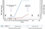
Figure. Tick-borne encephalitis in pregnant woman and long-term sequelae showing relevant clinical and laboratory findings, including TBEV antibody kinetics in serum samples. TBEV IgM (blue curve) and IgG (red curve) were...
On day 3, TBEV serologic results were positive for IgM and negative for IgG (Figure). The patient progressed to severe hyperactive delirium, requiring sedation and intubation. After a second lumbar puncture, results of reverse transcription PCR testing of cerebrospinal fluid (CSF) was positive for TBEV (Figure). A second MRI showed signs of diffuse leptomeningitis with deep cerebral nuclei involvement (Appendix Figure). At cessation of sedation (day 7), the patient remained in a coma. Iterative TBEV serologic results showed appearance of specific IgG (Figure).
At admission (day 0), lumbar puncture showed pleocytosis (70 cells/mm3) and 55 mg/dL of protein, and all virologic molecular tests results and bacterial culture results were negative. Lyme borreliosis serologic test results were negative. Results of magnetic resonance imaging (MRI) of the brain were unremarkable. Faced with a worsening of her condition, she was transferred to the intensive care unit 2 days later and showed aseptic meningoencephalitis. Given her recent history of travel, we tested for West Nile virus, dengue virus, Zika virus, Toscana virus, and chikungunya virus; all results were negative. An autoimmune etiology was ruled out by biologic testing.
At the beginning of August 2020, the patient was transferred to Charité Universitätsmedizin in Berlin, Germany. The next 2 MRIs, performed in September and November 2020, showed progression to deep cerebral nuclei and thalamic hemorrhagic transformation and cerebral atrophy (Appendix Figure). She was discharged to a neurologic rehabilitation center after 85 days of hospitalization and had tetraparesis and polyradiculitis. A tracheostomy and a gastrostomy were performed. After intensive rehabilitation, the patient showed slow and minor clinical improvement. She was not vaccinated against TBE and did not recall either a tick bite or consumption of raw milk products. All uterine ultrasounds performed during her hospitalization showed development of the fetus on schedule. The patient gave birth to a healthy boy by cesarean delivery at term.
Six previous cases of TBEV infection occurring during pregnancy have been published (2,3). For this case, as well as for 2 previously reported cases, pregnancy proceeded normally despite severe maternal infection (4). However, for 2 cases reported in 1966 (3), premature birth and fetal or neonatal intracranial hemorrhage occurred after the mother was infected. Although vertical transmission is known to occur with other arboviruses, such as Zika virus, to date, it has not been demonstrated for TBEV in humans (4) and has only been described in some animal models (5,6). Transplacental transmission seems unlikely because of the barrier function of the placenta and the short time of TBEV viremia in natural infection (1).
Pregnancy-associated changes in the immune system probably influenced the critical state of the patient. Usually, during the phase involving central nervous system symptoms, specific TBEV antibodies appear in blood or CSF samples, but viral RNA cannot be detected in those biologic fluids. TBEV RNA is rarely detected in CSF samples, as for our patient, corresponding to severe or fatal cases occurring in immunosuppressed patients (7,8). Relative to pregnancy-related immunotolerance, this patient also showed development of a delayed humoral immune response to TBEV because the first serologic results were negative at the onset of clinical central nervous system disease (1,7).
Cellular immune response is also required for the clearance of TBEV infection (1,6,9,10). We did not explore the cell-mediated response of our patient, but it was potentially also weakened by her pregnancy condition, which could explain the prolonged viral replication in CSF.
Concordant with the severe disease progression of this patient, iterative MRI showed cerebral meningo-radiculoencephalitis evolving to deep cerebral nuclei and thalamic hemorrhagic transformation and cerebral atrophy. Abnormalities on brain MRI are reported in only 20% of TBE patients (7).
As for all previously reported cases, this patient was not vaccinated against TBE. However, an effective vaccine is available and not contraindicated during pregnancy.
Further research is warranted to assess the course of TBEV infection during pregnancy. In this context, our case study offers relevant information and highlights the need for vaccination against TBE in disease-endemic areas.
Dr. Velay is an assistant professor in the Department of Virology at Strasbourg University Hospital, Strasbourg, France. Her primary research interest is vectorborne diseases with a focus on tick-borne encephalitis.
References
- Velay A, Paz M, Cesbron M, Gantner P, Solis M, Soulier E, et al. Tick-borne encephalitis virus: molecular determinants of neuropathogenesis of an emerging pathogen. Crit Rev Microbiol. 2019;45:472–93. DOIPubMedGoogle Scholar
- Schwaiger M, Cassinotti P. Development of a quantitative real-time RT-PCR assay with internal control for the laboratory detection of tick borne encephalitis virus (TBEV) RNA. J Clin Virol. 2003;27:136–45. DOIPubMedGoogle Scholar
- Weinmayr LM, Kanz D, Eckenweiler M, Bormann T, Huzly D, Bardutzky J, et al. Acute tick-borne encephalitis during pregnancy - An alarming combination. Ticks Tick Borne Dis. 2020;11:
101512 . DOIPubMedGoogle Scholar - Divé I, Veje M, Dobler G, Bergström T, Buxmann H, Paul B, et al. Tick-borne encephalitis virus (TBEV) infection in pregnancy: Absence of virus transmission to the fetuses despite severe maternal disease - A case study. Ticks Tick Borne Dis. 2020;11:
101491 . DOIPubMedGoogle Scholar - Charlier C, Beaudoin M-C, Couderc T, Lortholary O, Lecuit M. Arboviruses and pregnancy: maternal, fetal, and neonatal effects. Lancet Child Adolesc Health. 2017;1:134–46. DOIPubMedGoogle Scholar
- Bakhvalova VN, Potapova OF, Panov VV, Morozova OV. Vertical transmission of tick-borne encephalitis virus between generations of adapted reservoir small rodents. Virus Res. 2009;140:172–8. DOIPubMedGoogle Scholar
- Taba P, Schmutzhard E, Forsberg P, Lutsar I, Ljøstad U, Mygland Å, et al. EAN consensus review on prevention, diagnosis and management of tick-borne encephalitis. Eur J Neurol. 2017;24:1214–e61. DOIPubMedGoogle Scholar
- Lipowski D, Popiel M, Perlejewski K, Nakamura S, Bukowska-Osko I, Rzadkiewicz E, et al. A cluster of fatal tick-borne encephalitis virus infection in organ transplant setting. J Infect Dis. 2017;215:896–901. DOIPubMedGoogle Scholar
- Silasi M, Cardenas I, Kwon J-Y, Racicot K, Aldo P, Mor G. Viral infections during pregnancy. Am J Reprod Immunol. 2015;73:199–213. DOIPubMedGoogle Scholar
- Blom K, Cuapio A, Sandberg JT, Varnaite R, Michaëlsson J, Björkström NK, et al. Cell-mediated immune responses and immunopathogenesis of human tick-borne encephalitis virus-infection. Front Immunol. 2018;9:2174. DOIPubMedGoogle Scholar
Figure
Cite This ArticleOriginal Publication Date: February 14, 2023
Table of Contents – Volume 29, Number 3—March 2023
| EID Search Options |
|---|
|
|
|
|
|
|
Please use the form below to submit correspondence to the authors or contact them at the following address:
Aurélie Velay, Virology Laboratory, University Hospital of Strasbourg, Strasbourg F-67000, France
Top