Volume 29, Number 4—April 2023
Dispatch
Ocular Trematodiasis in Children, Sri Lanka
Figure 1

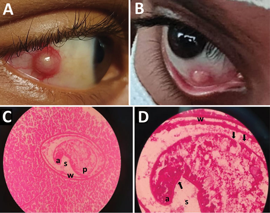
Figure 1. Trematode infection in the eyes of 2 pediatric patients in a study of ocular trematodiasis in children, Sri Lanka. A, B) Episcleral nodules found in eyes of 2 male pediatric patients. C, D) Metacercarial stage of a trematode in hematoxylin/eosin-stained tissue section of an excised episcleral nodule from a 12-year old boy. a, anterior end; p, posterior end; s, space between the larva and cyst wall; w, double layer cyst wall. Arrows in panel D indicate possible spines on surface tegument. Original magnification ×40 for panel C, ×100 for panel D.
Page created: January 25, 2023
Page updated: March 21, 2023
Page reviewed: March 21, 2023
The conclusions, findings, and opinions expressed by authors contributing to this journal do not necessarily reflect the official position of the U.S. Department of Health and Human Services, the Public Health Service, the Centers for Disease Control and Prevention, or the authors' affiliated institutions. Use of trade names is for identification only and does not imply endorsement by any of the groups named above.