Skip directly to site content Skip directly to page options Skip directly to A-Z link Skip directly to A-Z link Skip directly to A-Z link
Volume 29, Number 6—June 2023
Dispatch

Replication of Novel Zoonotic-Like Influenza A(H3N8) Virus in Ex Vivo Human Bronchus and Lung

Kenrie P.Y. Hui, John C.W. Ho, Ka-Chun Ng, Samuel M.S. Cheng, Ko-Yung Sit, Timmy W.K. Au, Leo L.M. Poon, John M. Nicholls, Malik Peiris, and Michael C.W. ChanComments to Author 
Author affiliations: The University of Hong Kong School of Public Health, Hong Kong, China (K.P.Y. Hui, J.C.W. Ho, K.-C. Ng, S.M.S. Cheng, L.L.M. Poon, M. Peiris, M.C.W. Chan); Centre for Immunology and Infection, Hong Kong (K.P.Y. Hui, J.C.W. Ho, L.L.M. Poon, M. Peiris, M.C.W. Chan); The University of Hong Kong Division of Cardiothoracic Surgery, Hong Kong (K.-Y. Sit, T.W.K. Au); The University of Hong Kong School of Clinical Medicine, Hong Kong (J.M. Nicholls)

Main Article

Figure 1

Comparative replication competence of zoonotic-like influenza A(H3N8) viruses isolated from chicken and other human and avian viruses in ex vivo cultures of human bronchus and lung tissue. Viral M segment RNA copies (A, B) and viral titers (E, F) in culture supernatants were collected at 1, 24, and 48 hours postinfection with H9N2/Y280, pH1N1, avH3N8/MP16, novel H3N8, or H5N1/483 viruses and measured by quantitative reverse transcription PCR (A, B) and TCID50 (E, F). C, D) Viral load from panels A and B by virus strain. G, H) Viral titers from panels E and F by virus strain. Data are geometric mean +SD. Statistical analysis was performed using 2-way (A, B, E, F)or 1-way (C, D, G, H) analysis of variance followed by Tukey posttest; p<0.05 was considered to be statistically significant. Detailed information on viruses used in study is provided in the Appendix. AUC, area under the curve; M, matrix; TCID50, 50% tissue culture infectious dose.

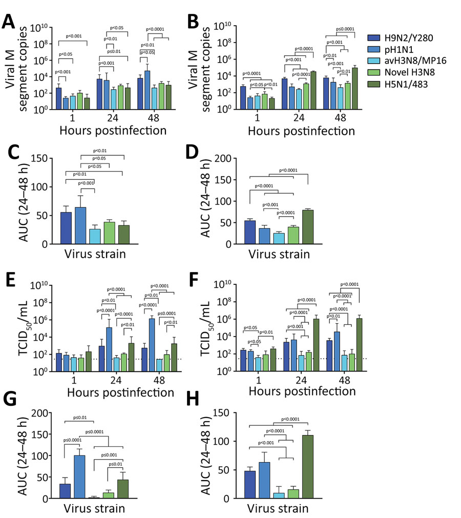
Figure 1. Comparative replication competence of zoonotic-like influenza A(H3N8) viruses isolated from chicken and other human and avian viruses in ex vivo cultures of human bronchus and lung tissue. Viral M segment RNA copies (A, B) and viral titers (E, F) in culture supernatants were collected at 1, 24, and 48 hours postinfection with H9N2/Y280, pH1N1, avH3N8/MP16, novel H3N8, or H5N1/483 viruses and measured by quantitative reverse transcription PCR (A, B) and TCID50 (E, F). C, D) Viral load from panels A and B by virus strain. G, H) Viral titers from panels E and F by virus strain. Data are geometric mean +SD. Statistical analysis was performed using 2-way (A, B, E, F)or 1-way (C, D, G, H) analysis of variance followed by Tukey posttest; p<0.05 was considered to be statistically significant. Detailed information on viruses used in study is provided in the Appendix. AUC, area under the curve; M, matrix; TCID50, 50% tissue culture infectious dose.

Main Article

Page created: March 29, 2023
Page updated: May 17, 2023
Page reviewed: May 17, 2023
The conclusions, findings, and opinions expressed by authors contributing to this journal do not necessarily reflect the official position of the U.S. Department of Health and Human Services, the Public Health Service, the Centers for Disease Control and Prevention, or the authors' affiliated institutions. Use of trade names is for identification only and does not imply endorsement by any of the groups named above.
file_external