Volume 29, Number 8—August 2023
Dispatch
Candidatus Neoehrlichia mikurensis Infection in Patient with Antecedent Hematologic Neoplasm, Spain1
Cite This Article
Citation for Media
Abstract
We report a confirmed case of Candidatus Neoehrlichia mikurensis infection in a woman in Spain who had a previous hematologic malignancy. Candidatus N. mikurensis infections should be especially suspected in immunocompromised patients who exhibit persistent fever and venous thrombosis, particularly if they live in environments where ticks are prevalent.
Candidatus Neoehrlichia mikurensis is an α1-proteobacterium (family Anaplasmataceae) transmitted by Ixodes spp. ticks. Although previously described in ticks and mammals in Europe and Asia, the species name was derived from a report in 2004 from Mikura Island, Japan, where the bacterium was found in endothelial cells from rat (Rattus norvegicus) spleens and in Ixodes ovatus ticks (1). In 2010, Candidatus N. mikurensis was identified as a human pathogen in Sweden (2). Since then, several case series and individual cases of patients with Candidatus N. mikurensis infections have been described, mainly in persons who were immunosuppressed because of hematologic neoplasms, splenectomies, or immunosuppressive drug treatment (3–9). However, Candidatus N. mikurensis can cause disease (neoehrlichiosis) in immunocompetent persons or cause asymptomatic infections (10,11). In 2019, Candidatus N. mikurensis was cultured in tick cell lines and infection was transferred to human endothelial cells derived from skin microvasculature and pulmonary arteries, demonstrating endothelial cell tropism. Tropism partly explains the clinical spectrum caused by the bacterium, usually consisting of persistent and recurrent fever and thrombosis and vasculitis with or without erysipelas-like skin lesions (12). In Spain, Candidatus N. mikurensis was found in Ixodes ricinus ticks removed from cows in 2013, but the bacterium was not detected in humans (13). We describe a case of Candidatus N. mikurensis infection in an immunocompromised patient from Asturias in northern Spain.
In September 2020, stage IV-B germinal center diffuse large B-cell lymphoma was diagnosed in a splenectomy specimen from a 68-year-old woman. She completed first-line treatment with rituximab plus cyclophosphamide, doxorubicin, vincristine, and prednisone and achieved complete remission. On June 21, 2021 (≈5 months after lymphoma treatment had ended), she experienced arthromyalgia, anorexia, night sweats, and vespertine fever. Her family physician began treatment with metamizole and cefuroxime at usual doses because of urine sediment alterations. Several days later, deep vein thrombosis developed in her right leg. Because of her previous malignancy and treatment, she was attended at her hospital’s hematology service. She was slightly anemic (hemoglobin 11.7 g/dL, reference range 12–16 g/dL) and had leukopenia (2.28 × 10³ leukocytes/µL, reference range 4–14 × 103 leukocytes/µL) and a low neutrophil count (0.4 × 10³ neutrophils/µL, reference range 1.8–8.5 × 10³ neutrophils/µL). C-reactive protein level was elevated (62 mg/L, reference range <10 mg/L), hyponatremia was present (133 mmol Na/L, reference range 135–145 mmol Na/L), and high levels of ferritin (536 µg /L, reference range 20–200 µg/L) and β2 microglobulin (8.50 mg/L, reference range 0.8–2.4 mg/L) were observed. Other measured hematologic and biochemical parameters, including procalcitonin, were within reference ranges. Other analyses, such as antinuclear antibody testing, blood and urine cultures, and serologic assays against Coxiella burnetii, herpes virus, cytomegalovirus, and Epstein-Barr virus, did not indicate acute infection. A chest radiograph and computed tomography scan and an abdominal ultrasound did not reveal pertinent abnormalities. Recurrence of lymphoma was suspected, and a positron emission tomography/computed tomography scan showed diffuse and homogeneous bone marrow hypermetabolism without evidence of neoplastic activity at other levels.
Empirical treatment was begun with piperacillin/tazobactam and granulocyte colony stimulating factor at conventional doses; 1 week later, the patient had recovered from leukopenia, but fever persisted. A bone marrow biopsy, which did not show neoplastic infiltration or alterations in hematopoietic cells, was performed and processed for different microbiologic tests. A possible tick-related infection was suspected because the patient lived in an area endemic for Lyme disease and other tickborne diseases. The patient recalled having suffered a tick bite 20 days before onset of symptoms. A bone marrow DNA extract and serum sample collected during the acute infection phase (August 2021) were sent to the Special Pathogens Laboratory, Center for Rickettsioses and Arthropod-Borne Diseases, at San Pedro University Hospital–Center for Biomedical Research of La Rioja in Logroño, Spain, to screen for Candidatus N. mikurensis by using PCR and Anaplasma phagocytophilum by using PCR and immunofluorescence assays.
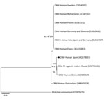
We performed PCR targeting the panbacterial 16S rRNA gene, fragments of 16S rRNA gene from Anaplasmataceae (designated as 16S rRNA-EHR), groEL from Candidatus N. mikurensis, and msp2 from A. phagocytophilum (Table 1). We detected PCR amplicons of the expected sizes for groEL and panbacteria and family-specific 16S rRNA in bone marrow and acute phase serum samples; nucleotide sequences corresponded to Candidatus N. mikurensis. The groEL amplicon (1,232 bp) showed the highest (99.3%) sequence similarity with that of Candidatus N. mikurensis from a wild rodent (Microtus agrestis) from Siberia in Russia (GenBank accession no. MN701626) but differed from other highly conserved sequences from Siberia and the Far East; the sequence was 98.8% identical to Candidatus N. mikurensis found in Ixodes ricinus ticks from Spain (13) (Table 2). We constructed a phylogenetic tree for groEL sequences by using the maximum likelihood method (Figure). We found no differences for the 16S rRNA-EHR sequence (306 bp). The panbacteria 16S rRNA sequence (available upon request from the authors) showed 3–27 mismatches with the 16S rRNA from Candidatus N. mikurensis. We did not detect A. phagocytophilum by PCR in the acute samples. We deposited nucleotide sequences of groEL and 16S rRNA genes generated in this study in GenBank under accession nos. OQ579033 (groEL) and OQ581737 (16S rRNA).
On the basis of PCR results, the patient was treated with doxycycline (100 mg 2×/d for 3 wk), and fever disappeared after 72 hours. Neutropenia was attributed to the intake of metamizole for symptom control. However, another case of doxycycline-treated Candidatus N. mikurensis infection associated with neutropenia has been reported (8). EDTA-anticoagulated blood and serum specimens were collected 4 (December 2021) and 6 (February 2022) months after onset of the acute infection phase, and we screened for Candidatus N. mikurensis at the Center for Rickettsioses and Arthropod-Borne Diseases, as previously described. We detected Candidatus N. mikurensis DNA in blood collected at 4 months but not in serum. The patient was healthy and blood test results did not show abnormalities at that time. Follow-up PCR of specimens collected at 6 months yielded negative results (Table 2). We did not detect IgG against A. phagocytophilum.
We report a confirmed case of Candidatus N. mikurensis infection in Spain, detected in human bone marrow aspirate, serum, and EDTA-blood samples, that was no longer detected months after completing antimicrobial drug treatment. A broad clinical spectrum of tickborne diseases is found in Spain. Human cases of Lyme borreliosis, Mediterranean spotted fever, and other tickborne rickettsioses have been described, including Dermacentor tick–borne necrosis erythema lymphadenopathy, Rickettsia sibirica mongolitimonae infection, R. massiliae infection, R. aeschlimannii infection, babesiosis, human anaplasmosis, tularemia, Borrelia hispanica relapsing fever, tick paralysis, Crimean-Congo hemorrhagic fever, and α-gal syndrome or other allergic reactions (14). Since we discovered Candidatus N. mikurensis in I. ricinus ticks in Spain (13), we have conducted surveillance of this bacterium. Candidatus N. mikurensis should be considered a potential cause of persistent fever and venous thrombosis in patients with hematologic malignancies who live in environments where ticks are prevalent. Candidatus N. mikurensis infections should be particularly suspected in patients who are immunosuppressed but also should be considered in patients with other vascular conditions who are not immunocompromised (15).
Dr. González-Carmona is a hematologist at Hospital de Jarrio in Asturias, Spain. Her research interests focus on opportunistic infections in cancer patients.
Acknowledgments
We thank Sonia Santibáñez, Ana M. Palomar, Ignacio Ruiz-Arrondo, and Paula Santibáñez for technical assistance.
This study was partially funded by Ministerio de Ciencia, Innovación y Universidades, Instituto de Salud Carlos III through the Spanish Network for Research in Infectious Diseases (RD16/0016/0013) (https://www.reipi.org) and co-funded by the European Regional Development Fund, A way to achieve Europe, ERDF.
References
- Kawahara M, Rikihisa Y, Isogai E, Takahashi M, Misumi H, Suto C, et al. Ultrastructure and phylogenetic analysis of ‘Candidatus Neoehrlichia mikurensis’ in the family Anaplasmataceae, isolated from wild rats and found in Ixodes ovatus ticks. Int J Syst Evol Microbiol. 2004;54:1837–43. DOIPubMedGoogle Scholar
- Welinder-Olsson C, Kjellin E, Vaht K, Jacobsson S, Wennerås C. First case of human “Candidatus Neoehrlichia mikurensis” infection in a febrile patient with chronic lymphocytic leukemia. J Clin Microbiol. 2010;48:1956–9. DOIPubMedGoogle Scholar
- von Loewenich FD, Geissdörfer W, Disqué C, Matten J, Schett G, Sakka SG, et al. Detection of “Candidatus Neoehrlichia mikurensis” in two patients with severe febrile illnesses: evidence for a European sequence variant. J Clin Microbiol. 2010;48:2630–5. DOIPubMedGoogle Scholar
- Pekova S, Vydra J, Kabickova H, Frankova S, Haugvicova R, Mazal O, et al. Candidatus Neoehrlichia mikurensis infection identified in 2 hematooncologic patients: benefit of molecular techniques for rare pathogen detection. Diagn Microbiol Infect Dis. 2011;69:266–70. DOIPubMedGoogle Scholar
- Maurer FP, Keller PM, Beuret C, Joha C, Achermann Y, Gubler J, et al. Close geographic association of human neoehrlichiosis and tick populations carrying “Candidatus Neoehrlichia mikurensis” in eastern Switzerland. J Clin Microbiol. 2013;51:169–76. DOIPubMedGoogle Scholar
- Grankvist A, Andersson PO, Mattsson M, Sender M, Vaht K, Höper L, et al. Infections with the tick-borne bacterium “Candidatus Neoehrlichia mikurensis” mimic noninfectious conditions in patients with B cell malignancies or autoimmune diseases. Clin Infect Dis. 2014;58:1716–22. DOIPubMedGoogle Scholar
- Andréasson K, Jönsson G, Lindell P, Gülfe A, Ingvarsson R, Lindqvist E, et al. Recurrent fever caused by Candidatus Neoehrlichia mikurensis in a rheumatoid arthritis patient treated with rituximab. Rheumatology (Oxford). 2015;54:369–71. DOIPubMedGoogle Scholar
- Lenart M, Simoniti M, Strašek-Smrdel K, Špik VC, Selič-Kurinčič T, Avšič-Županc T. Case report: first symptomatic Candidatus Neoehrlichia mikurensis infection in Slovenia. BMC Infect Dis. 2021;21:579. DOIPubMedGoogle Scholar
- Boyer PH, Baldinger L, Degeilh B, Wirth X, Kamdem CM, Hansmann Y, et al. The emerging tick-borne pathogen Neoehrlichia mikurensis: first French case series and vector epidemiology. Emerg Microbes Infect. 2021;10:1731–8. DOIPubMedGoogle Scholar
- Li H, Jiang JF, Liu W, Zheng YC, Huo QB, Tang K, et al. Human infection with Candidatus Neoehrlichia mikurensis, China. Emerg Infect Dis. 2012;18:1636–9. DOIPubMedGoogle Scholar
- Portillo A, Santibáñez P, Palomar AM, Santibáñez S, Oteo JA. ‘Candidatus Neoehrlichia mikurensis’ in Europe. New Microbes New Infect. 2018;22:30–6. DOIPubMedGoogle Scholar
- Wass L, Grankvist A, Bell-Sakyi L, Bergström M, Ulfhammer E, Lingblom C, et al. Cultivation of the causative agent of human neoehrlichiosis from clinical isolates identifies vascular endothelium as a target of infection. Emerg Microbes Infect. 2019;8:413–25. DOIPubMedGoogle Scholar
- Palomar AM, García-Álvarez L, Santibáñez S, Portillo A, Oteo JA. Detection of tick-borne ‘Candidatus Neoehrlichia mikurensis’ and Anaplasma phagocytophilum in Spain in 2013. Parasit Vectors. 2014;7:57. DOIPubMedGoogle Scholar
- Portillo A, Ruiz-Arrondo I, Oteo JA. Arthropods as vectors of transmissible diseases in Spain. [Engl Ed]. Med Clin (Engl Ed). 2018;151:450–9. DOIPubMedGoogle Scholar
- Höper L, Skoog E, Stenson M, Grankvist A, Wass L, Olsen B, et al. Vasculitis due to Candidatus Neoehrlichia mikurensis: a cohort study of 40 Swedish patients. Clin Infect Dis. 2021;73:e2372–8. DOIPubMedGoogle Scholar
Figure
Tables
Cite This Article1Data from this study were presented at the joint LXIV National Conference of the Spanish Society of Hematology and Hemotherapy, XXXVIII National Conference of the Spanish Society of Thrombosis and Hemostasis, and 38th World Congress of the International Society of Hematology; October 6–8, 2022; Barcelona, Spain; and International Intracellular Bacteria Meeting; August 23–26, 2022; Lausanne, Switzerland.
2These first authors contributed equally to this article.
3Current affiliation: Hospital Universitario San Agustín, Asturias, Spain.
Table of Contents – Volume 29, Number 8—August 2023
| EID Search Options |
|---|
|
|
|
|
|
|

Please use the form below to submit correspondence to the authors or contact them at the following address:
José A. Oteo, Departamento de Enfermedades Infecciosas, Hospital Universitario San Pedro-CIBIR, Centro de Rickettsiosis y Enfermedades Transmitidas por Artrópodos Vectores, C/Piqueras 98, 26006 Logroño, La Rioja, Spain
Top