Volume 29, Number 9—September 2023
Research
Interspecies Transmission of Swine Influenza A Viruses and Human Seasonal Vaccine-Mediated Protection Investigated in Ferret Model
Figure 6
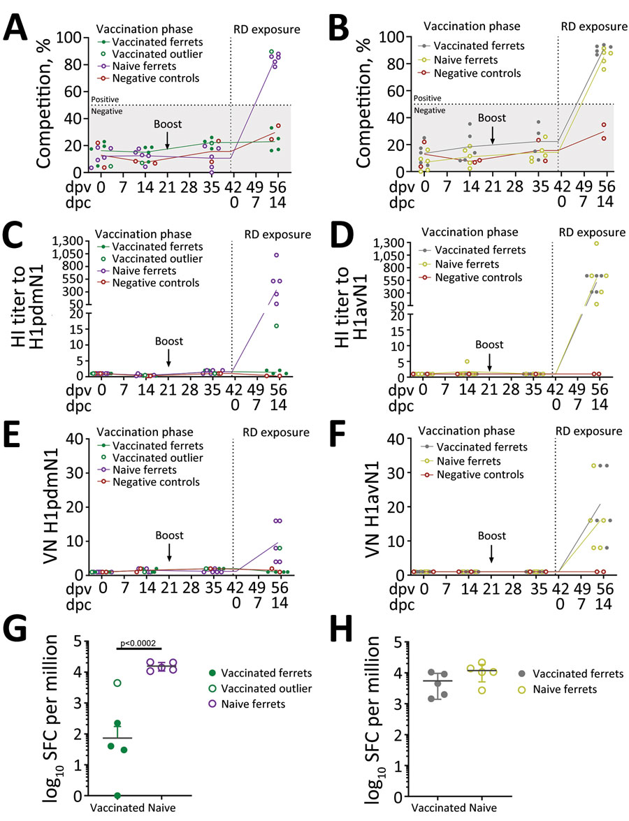
Figure 6. Immune parameters assessed in naive and vaccinated ferrets before and after exposure to pigs infected with influenza A viruses A/swine/England/1353/2009 (H1pdmN1, panels A, C, E, and G) or A/Pavia/65/2016 (H1avN1, panels B, D, F, and H). Data from a single outlier, a vaccinated ferret exposed to the H1pdmN1 virus, were excluded from analysis but are shown. Negative control ferrets (n = 2) were not vaccinated or exposed to infectious virus. Specific humoral responses were assessed longitudinally in serum. Antibody titers detected by NP competition ELISA (A, B) are expressed as competition percentage and considered negative if <50% (gray area). Competition percentage was calculated as (1 – sample/negative) × 100. HI (C, D) and VN (E, F) were determined using the homologous virus for each group. Both HI and VN titers are normalized to the individual prevaccination titers (0 dpv). ELISpot analysis (G, H) evaluated the number of interferon-γ–producing peripheral blood mononuclear cells induced by 18-mer nucleoprotein peptides, represented as per 1 million, at 14 dpc (RD exposure). dpv, days postvaccination; dpc, days postcontact; HI, hemagglutination inhibition; NP, nucleoprotein; RD, respiratory droplets; SPC, spot-forming cells; VN, virus neutralization.