Volume 30, Number 12—December 2024
Research
Autochthonous Blastomyces dermatitidis, India
Figure 1

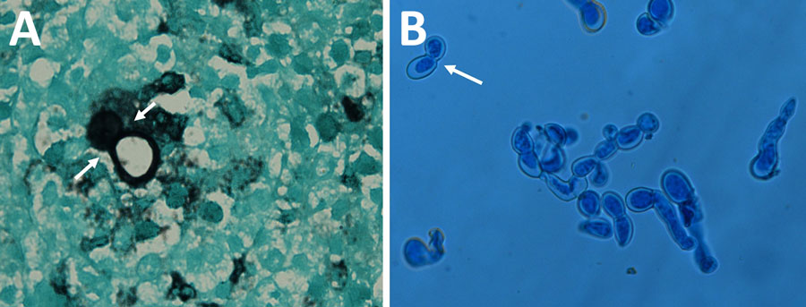
Figure 1. Autochthonous Blastomyces dermatitidis biopsy and culture findings from a patient in India. Microscopic characteristics are consistent with Blastomyces dermatitidis. A) Gomori’s methenamine silver–stained section of a subcutaneous nodule biopsy obtained from sternum region showing a large, thick-walled broad-based budding yeast cell (white arrows) typical of Blastomyces. B) Lactophenol cotton blue mount of the 6-day-old yeast form on pea seed agar at 37°C, showing numerous large thick-walled and broad-based budding yeast cells (white arrow). Original magnification is ×100.
Page created: November 13, 2024
Page updated: November 26, 2024
Page reviewed: November 26, 2024
The conclusions, findings, and opinions expressed by authors contributing to this journal do not necessarily reflect the official position of the U.S. Department of Health and Human Services, the Public Health Service, the Centers for Disease Control and Prevention, or the authors' affiliated institutions. Use of trade names is for identification only and does not imply endorsement by any of the groups named above.