Volume 30, Number 12—December 2024
Research
Autochthonous Blastomyces dermatitidis, India
Figure 2

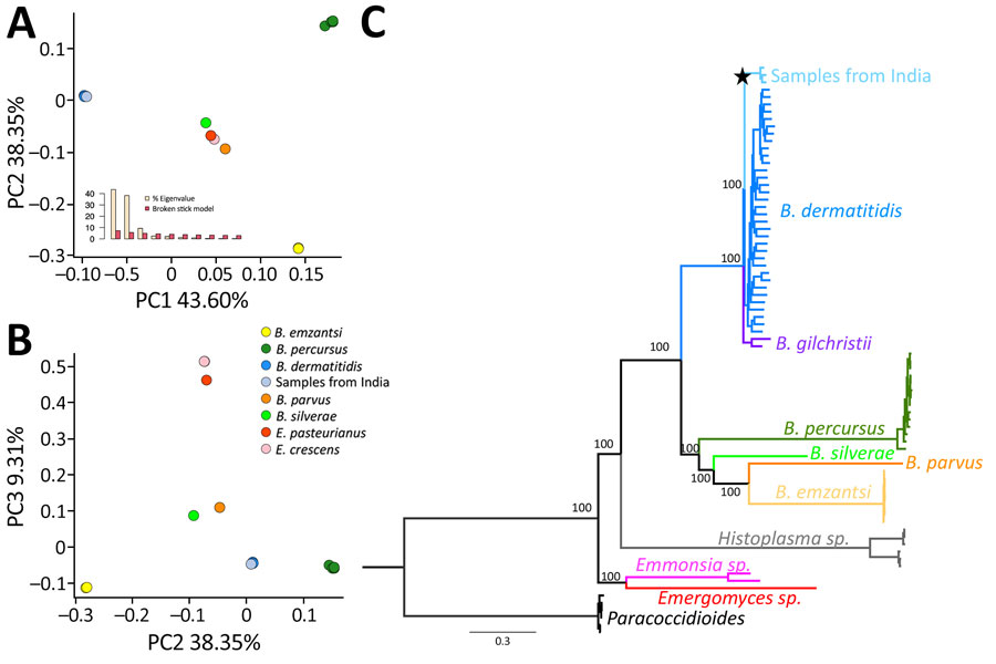
Figure 2. Genetic differentiation between 3 Blastomyces dermatitidis from India and other dimorphic fungi. A) PC analyses showing the 3 isolates from India (light blue) clustering with B. dermatitidis (dark blue) in PC 1 and PC2. The broken stick model (inset) demonstrates the first 3 PCs are significant. B) PC analyses showing the 3 isolates from India (light blue) clustering with B. dermatitidis (dark blue) in PC 32 and PC 3. C) Rooted phylogram for autochthonous B. dermatitidis isolates from India and the genetic relationships with other Blastomyces fungi. A maximum-likelihood tree derived from genomewide concatenated markers using the B. dermatitidis reference genome suggests a close phylogenetic relationship between B. dermatitidis and the 3 isolates from India. The star shows the node leading to the B. dermatitidis lineage from India. Scale bar represents the number of substitutions per site. PC, principal component.