Volume 30, Number 2—February 2024
Synopsis
Parechovirus A Circulation and Testing Capacities in Europe, 2015–2021
Figure 4

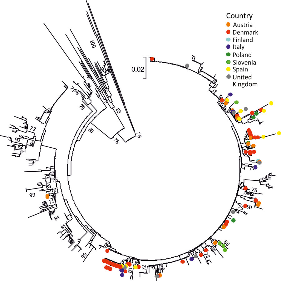
Figure 4. Phylogenetic analysis of the VP3/VP1 region of PeV-3 sequences, Europe, 2015–2021. Neighbor-joining phylogenetic tree of the VP3/VP1 junction region obtained from the study samples (n = 106) is labeled by country of sample origin and compared with 630 available sequences spanning the analyzed region from GenBank. The tree was constructed using MEGA 7 (17) using Jukes-Cantor corrected distances, with bootstrap resampling; branches showing 70% or greater supports were labeled. Scale bar indicates substitutions per site. A maximum-likelihood analysis of the same sequence dataset is provided in Appendix Figure 2. PeV, parechovirus type; VP, viral protein.
References
- Harvala H, Wolthers KC, Simmonds P. Parechoviruses in children: understanding a new infection. Curr Opin Infect Dis. 2010;23:224–30. DOIPubMedGoogle Scholar
- Wang CYT, Ware RS, Lambert SB, Mhango LP, Tozer S, Day R, et al. Parechovirus A infections in healthy Australian children during the first 2 years of life: a community-based longitudinal birth cohort study. Clin Infect Dis. 2020;71:116–27. DOIPubMedGoogle Scholar
- Olijve L, Jennings L, Walls T. Human parechovirus: an increasingly recognized cause of sepsis-like illness in young infants. Clin Microbiol Rev. 2017;31:e00047–17.DOIPubMedGoogle Scholar
- Harvala H, Robertson I, Chieochansin T, McWilliam Leitch EC, Templeton K, Simmonds P. Specific association of human parechovirus type 3 with sepsis and fever in young infants, as identified by direct typing of cerebrospinal fluid samples. J Infect Dis. 2009;199:1753–60. DOIPubMedGoogle Scholar
- Wolthers KC, Benschop KS, Schinkel J, Molenkamp R, Bergevoet RM, Spijkerman IJ, et al. Human parechoviruses as an important viral cause of sepsislike illness and meningitis in young children. Clin Infect Dis. 2008;47:358–63. DOIPubMedGoogle Scholar
- Harvala H, Calvert J, Van Nguyen D, Clasper L, Gadsby N, Molyneaux P, et al. Comparison of diagnostic clinical samples and environmental sampling for enterovirus and parechovirus surveillance in Scotland, 2010 to 2012. Euro Surveill. 2014;19:20772. DOIPubMedGoogle Scholar
- Zell R, Delwart E, Gorbalenya AE, Hovi T, King AMQ, Knowles NJ, et al.; Ictv Report Consortium. ICTV virus taxonomy profile: Picornaviridae. J Gen Virol. 2017;98:2421–2. DOIPubMedGoogle Scholar
- Harvala H, McLeish N, Kondracka J, McIntyre CL, McWilliam Leitch EC, Templeton K, et al. Comparison of human parechovirus and enterovirus detection frequencies in cerebrospinal fluid samples collected over a 5-year period in edinburgh: HPeV type 3 identified as the most common picornavirus type. J Med Virol. 2011;83:889–96. DOIPubMedGoogle Scholar
- Harvala H, Robertson I, McWilliam Leitch EC, Benschop K, Wolthers KC, Templeton K, et al. Epidemiology and clinical associations of human parechovirus respiratory infections. J Clin Microbiol. 2008;46:3446–53. DOIPubMedGoogle Scholar
- Sasidharan A, Harrison CJ, Banerjee D, Selvarangan R. Emergence of parechovirus A4 central nervous system infections among infants in Kansas City, Missouri, USA. J Clin Microbiol. 2019;57:e01698–18. DOIPubMedGoogle Scholar
- Chamings A, Liew KC, Reid E, Athan E, Raditsis A, Vuillermin P, et al. An emerging human parechovirus type 5 causing sepsis-like illness in infants in Australia. Viruses. 2019;11:913. DOIPubMedGoogle Scholar
- Sridhar A, Karelehto E, Brouwer L, Pajkrt D, Wolthers KC. Parechovirus A pathogenesis and the enigma of genotype A-3. Viruses. 2019;11:1062. DOIPubMedGoogle Scholar
- EUSurvey. HPeV circulation in EU/EEA & UK, 2015–2021 [cited 2022 Mar 10]. https://ec.europa.eu/eusurvey/runner/HPeV_circulation_in_EU-EEA_UK_2015-2021
- Campbell I. Chi-squared and Fisher-Irwin tests of two-by-two tables with small sample recommendations. Stat Med. 2007;26:3661–75. DOIPubMedGoogle Scholar
- Edgar RC. MUSCLE: multiple sequence alignment with high accuracy and high throughput. Nucleic Acids Res. 2004;32:1792–7. DOIPubMedGoogle Scholar
- Simmonds P. SSE: a nucleotide and amino acid sequence analysis platform. BMC Res Notes. 2012;5:50. DOIPubMedGoogle Scholar
- Kumar S, Stecher G, Tamura K. MEGA7: Molecular Evolutionary Genetics Analysis version 7.0 for bigger datasets. Mol Biol Evol. 2016;33:1870–4. DOIPubMedGoogle Scholar
- Marchand S, Launay E, Schuffenecker I, Gras-Le Guen C, Imbert-Marcille BM, Coste-Burel M. Severity of parechovirus infections in infants under 3 months of age and comparison with enterovirus infections: A French retrospective study. Arch Pediatr. 2021;28:291–5. DOIPubMedGoogle Scholar
- Elling R, Böttcher S, du Bois F, Müller A, Prifert C, Weissbrich B, et al. Epidemiology of human parechovirus type 3 upsurge in 2 hospitals, Freiburg, Germany, 2018. Emerg Infect Dis. 2019;25:1384–8. DOIPubMedGoogle Scholar
- Linhares MI, Brett A, Correia L, Pereira H, Correia C, Oleastro M, et al. Parechovirus genotype 3 outbreak among young infants in Portugal. Acta Med Port. 2021;34:664–8. DOIPubMedGoogle Scholar
- Fischer TK, Midgley S, Dalgaard C, Nielsen AY. Human parechovirus infection, Denmark. Emerg Infect Dis. 2014;20:83–7. DOIPubMedGoogle Scholar
- Davis J, Fairley D, Christie S, Coyle P, Tubman R, Shields MD. Human parechovirus infection in neonatal intensive care. Pediatr Infect Dis J. 2015;34:121–4. DOIPubMedGoogle Scholar
- Centers for Disease Control and Prevention. National Enterovirus Surveillance System (NESS): surveillance data [cited 2023 Aug 28]. https://www.cdc.gov/surveillance/ness/surv-data.html
- Abedi GR, Watson JT, Nix WA, Oberste MS, Gerber SI. Enterovirus and parechovirus surveillance—United States, 2014–2016. MMWR Morb Mortal Wkly Rep. 2018;67:515–8. DOIPubMedGoogle Scholar
- Kadambari S, Harvala H, Simmonds P, Pollard AJ, Sadarangani M. Strategies to improve detection and management of human parechovirus infection in young infants. Lancet Infect Dis. 2019;19:e51–8. DOIPubMedGoogle Scholar
- Benschop KS, Schinkel J, Minnaar RP, Pajkrt D, Spanjerberg L, Kraakman HC, et al. Human parechovirus infections in Dutch children and the association between serotype and disease severity. Clin Infect Dis. 2006;42:204–10. DOIPubMedGoogle Scholar
- Piralla A, Furione M, Rovida F, Marchi A, Stronati M, Gerna G, et al. Human parechovirus infections in patients admitted to hospital in Northern Italy, 2008-2010. J Med Virol. 2012;84:686–90. DOIPubMedGoogle Scholar
- Harvala H, Griffiths M, Solomon T, Simmonds P. Distinct systemic and central nervous system disease patterns in enterovirus and parechovirus infected children. J Infect. 2014;69:69–74. DOIPubMedGoogle Scholar
- Esposito S, Rahamat-Langendoen J, Ascolese B, Senatore L, Castellazzi L, Niesters HG. Pediatric parechovirus infections. J Clin Virol. 2014;60:84–9. DOIPubMedGoogle Scholar
- van der Sanden S, de Bruin E, Vennema H, Swanink C, Koopmans M, van der Avoort H. Prevalence of human parechovirus in the Netherlands in 2000 to 2007. J Clin Microbiol. 2008;46:2884–9. DOIPubMedGoogle Scholar
- Nelson TM, Vuillermin P, Hodge J, Druce J, Williams DT, Jasrotia R, et al. An outbreak of severe infections among Australian infants caused by a novel recombinant strain of human parechovirus type 3. Sci Rep. 2017;7:44423. DOIPubMedGoogle Scholar
- Centers for Disease Control and Prevention. Emergency preparedness and response: recent reports of human parechovirus (PeV) in the United States—2022 [cited 2022 Jul 11]. https://emergency.cdc.gov/han/2022/han00469.asp
- South Carolina Department of Health and Environmental Control; South Carolina Health Alert Network. CDC health advisory: recent reports of human parechovirus (PeV) in the United States—2022 [cited 2022 Jul 13]. https://scdhec.gov/sites/default/files/media/document/10523-CHA-07-13-2022-PeV.pdf
- Victoria Department of Health. Human parechovirus type 3 in Victoria [cited 2022 Nov 7]. https://www.health.vic.gov.au/health-advisories/human-parechovirus-type-3-in-victoria
- Benschop KS, Albert J, Anton A, Andrés C, Aranzamendi M, Armannsdóttir B, et al. Re-emergence of enterovirus D68 in Europe after easing the COVID-19 lockdown, September 2021. Euro Surveill. 2021;26:
2100998 . DOIPubMedGoogle Scholar - Kolehmainen P, Jääskeläinen A, Blomqvist S, Kallio-Kokko H, Nuolivirta K, Helminen M, et al. Human parechovirus type 3 and 4 associated with severe infections in young children. Pediatr Infect Dis J. 2014;33:1109–13. DOIPubMedGoogle Scholar
- Piralla A, Perniciaro S, Ossola S, Giardina F, De Carli A, Bossi A, et al. Human parechovirus type 5 neurological infection in a neonate with a favourable outcome: A case report. Int J Infect Dis. 2019;89:175–8. DOIPubMedGoogle Scholar
Page created: December 13, 2023
Page updated: January 25, 2024
Page reviewed: January 25, 2024
The conclusions, findings, and opinions expressed by authors contributing to this journal do not necessarily reflect the official position of the U.S. Department of Health and Human Services, the Public Health Service, the Centers for Disease Control and Prevention, or the authors' affiliated institutions. Use of trade names is for identification only and does not imply endorsement by any of the groups named above.