Volume 30, Number 5—May 2024
Dispatch
Paranannizziopsis spp. Infection in Wild Vipers, Europe
Gaëlle Blanvillain , Fernando Martínez-Freiría, Joseph R. Hoyt, Jeffrey M. Lorch, and Albert Martinez-Silvestre
Figure 1
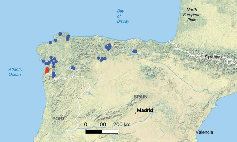
Figure 1. Spatial distribution of Seoane’s viper (Vipera seoanei) captures and detections of Paranannizziopsis sp. fungus in Spain and Portugal. Each dot represents an individual snake capture; overlapping points were slightly jittered for visualization. Blue dots represent snakes that tested negative by real-time PCR, and red dots represent snakes that tested positive by real-time PCR.
Page created: March 07, 2024
Page updated: April 24, 2024
Page reviewed: April 24, 2024
The conclusions, findings, and opinions expressed by authors contributing to this journal do not necessarily reflect the official position of the U.S. Department of Health and Human Services, the Public Health Service, the Centers for Disease Control and Prevention, or the authors' affiliated institutions. Use of trade names is for identification only and does not imply endorsement by any of the groups named above.