Volume 30, Number 5—May 2024
Dispatch
Paranannizziopsis spp. Infection in Wild Vipers, Europe
Gaëlle Blanvillain , Fernando Martínez-Freiría, Joseph R. Hoyt, Jeffrey M. Lorch, and Albert Martinez-Silvestre
Figure 2
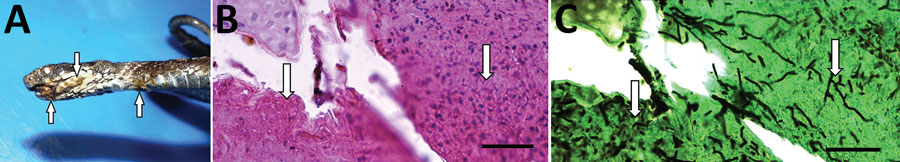
Figure 2. Seoane’s viper (Vipera seoanei) collected in Spain that was infected with Paranannizziopsis sp. fungus. A) Gross lesions in the mouth, on the lower jaw, and on the ventral areas of the body (arrows). B) Lightly stained hyphae (arrows)in section of epidermis stained with hematoxylin and eosin. Scale bar indicates 20 μm. C) Intralesional hyphae (arrows) in section of epidermis stained with the Grocott-Gomori methenamine silver method. Scale bar indicates 20 μm.
Page created: March 07, 2024
Page updated: April 24, 2024
Page reviewed: April 24, 2024
The conclusions, findings, and opinions expressed by authors contributing to this journal do not necessarily reflect the official position of the U.S. Department of Health and Human Services, the Public Health Service, the Centers for Disease Control and Prevention, or the authors' affiliated institutions. Use of trade names is for identification only and does not imply endorsement by any of the groups named above.