Skip directly to site content Skip directly to page options Skip directly to A-Z link Skip directly to A-Z link Skip directly to A-Z link

Disclaimer: Early release articles are not considered as final versions. Any changes will be reflected in the online version in the month the article is officially released.

Volume 30, Number 8—August 2024
Dispatch

Highly Pathogenic Avian Influenza Virus A(H5N1) Clade 2.3.4.4b Infection in Free-Ranging Polar Bear, Alaska, USA

Raphaela Stimmelmayr1Comments to Author , David Rotstein1, Mia Kim Torchetti, and Robert Gerlach
Author affiliations: North Slope Borough, Utqiagvik, Alaska, USA (R. Stimmelmayr); University of Alaska, Fairbanks, Alaska, USA (R. Stimmelmayr); Marine Mammal Pathology Services, Olney, Maryland, USA (D. Rotstein); US Department of Agriculture Animal and Plant Health Inspection Service, Ames, Iowa, USA (M.K. Torchetti); Alaska Department of Environmental Conservation, Anchorage, Alaska, USA (R. Gerlach)

Main Article

Figure

Histologic analysis of brain tissue from a dead free-ranging polar bear infected with highly pathogenic avian influenza virus A(H5N1) clade 2.3.4.4b, Alaska, USA. A) Hematoxylin and eosin staining of brain tissue section showing meningoencephalitis. Arrow indicates mixed inflammatory cells within and around blood vessels and hypertrophied vascular endothelial cells. Star indicates necrotic neurons and increased number of microglial cells within the parenchyma. Original magnification ×400. B) Arrow indicates influenza A virus within the neuronal perikaryon (red staining) observed by immunohistochemistry of formalin-fixed paraffin-embedded brain sections. Original magnification ×400.

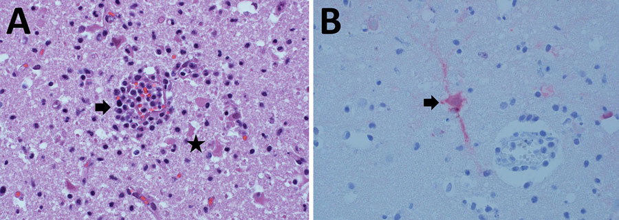
Figure. Histologic analysis of brain tissue from a dead free-ranging polar bear infected with highly pathogenic avian influenza virus A(H5N1) clade 2.3.4.4b, Alaska, USA. A) Hematoxylin and eosin staining of brain tissue section showing meningoencephalitis. Arrow indicates mixed inflammatory cells within and around blood vessels and hypertrophied vascular endothelial cells. Star indicates necrotic neurons and increased number of microglial cells within the parenchyma. Original magnification ×400. B) Arrow indicates influenza A virus within the neuronal perikaryon (red staining) observed by immunohistochemistry of formalin-fixed paraffin-embedded brain sections. Original magnification ×400.

Main Article

1These authors contributed equally to this article.

Page created: June 24, 2024
Page updated: June 28, 2024
Page reviewed: June 28, 2024
The conclusions, findings, and opinions expressed by authors contributing to this journal do not necessarily reflect the official position of the U.S. Department of Health and Human Services, the Public Health Service, the Centers for Disease Control and Prevention, or the authors' affiliated institutions. Use of trade names is for identification only and does not imply endorsement by any of the groups named above.
file_external