Volume 30, Number 9—September 2024
Research Letter
Molecular Confirmation of Taenia solium Taeniasis in Child, Timor-Leste
Cite This Article
Citation for Media
Abstract
We report a case of Taenia solium taeniasis in a 10-year-old child in Timor-Leste, confirmed by molecular analysis, suggesting T. solium transmission to humans is occurring in Timor-Leste. Proactive measures are needed to improve public understanding of prevalence, geographic spread, and health implications of human taeniasis and cysticercosis in Timor-Leste.
The pork tapeworm, Taenia solium, causes human taeniasis and cysticercosis, which are considerable health problems in many developing countries (1). In Southeast Asia, T. solium infections are considered endemic, but epidemiologic data remain scarce (2). We report a case of T. solium taeniasis in Timor-Leste, confirmed by molecular methods.
In March 2019, as part of routine monitoring by the Timor-Leste Ministry of Health’s national control program targeting soil-transmitted helminthiasis, in collaboration with the World Health Organization’s country office, 1,121 fecal samples from school children in Timor-Leste were examined by using the Kato-Katz method. Taenia spp. eggs were identified in 4 samples. Subsequently, we conducted home visits for each affected child and administered a single dose of 10 mg/kg praziquantel (Shin Poong Pharmaceutical Co. Ltd, https://shinpoong.co.kr). We were able to collect expelled worm segments on the same day of treatment from a 10-year-old girl residing in Dili, the capital of Timor-Leste. Throughout most of her life, the child had remained in good health and had not manifested symptoms indicative of human taeniasis. Also, she had not traveled outside of the country.
Figure 1
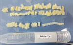
Figure 1. Proglottids of Taenia soliumcollected from a patient in Dili, Timor-Leste, in case study of molecular confirmation of taeniasis in a child. We collected the expelled worm segments from...
The retrieved worm segments exhibited a flat, creamy white appearance, aligning with the typical macroscopic characteristics associated with Taenia spp. (Figure 1). Microscopic analysis of the segments revealed ≈50 gravid, 20 mature, and 20 immature proglottids of T. solium.
Figure 2
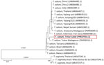
Figure 2. Phylogenetic analysis of the cox-1 gene in case study of Taenia soliumtaeniasis in a child, Timor-Leste. Evolutionary history was inferred by using the neighbor-joining method and...
To determine the species through molecular analysis, we isolated genomic DNA from 1 segment by using the DNeasy Blood & Tissue Kit (QIAGEN, https://www.qiagen.com), according to the manufacturer’s instructions. We performed PCR of genomic DNA to detect the parasite mitochondrial cox-1 gene that encodes cytochrome c oxidase subunit I (Appendix) (3). We purified the PCR products by using DNA Clean & Concentrator-5 (Zymo Research, https://www.zymoresearch.com), according to the manufacturer’s protocol. Sanger sequencing was subsequently performed by Bioneer Co., Ltd. (https://www.bioneer.co.kr), which used an ABI3730XL instrument (Applied Biosystems/Thermo Fisher Scientific, https://www.thermofisher.com). We deposited the derived sequence in GenBank (accession no. PP837933.1) and compared it with other cox-1 sequences in GenBank by using BLAST (https://blast.ncbi.nlm.nih.gov). The sequence showed 98.85%–100% identity with the T. solium mitochondrial cox-1 gene. We used cox-1 sequences for phylogenetic reconstruction (Figure 2; Appendix). We aligned DNA sequences by using ClustalW (http://www.clustal.org) and conducted evolutionary analyses by using MEGA11 (4). The sequence isolated in this study was shown to be most homologous with an isolate from Tulear (also known as Toliara), Madagascar (GenBank accession no. FM958316.1) (5). Consequently, molecular evaluation confirmed the infection was caused by T. solium.
We report documented human T. solium taeniasis in Timor-Leste, an area where previous records of the parasite have been nearly absent (6). Clinically diagnosed neurocysticercosis in persons from Timor have been reported in Australia and Indonesia, suggesting the presence of T. solium in Timor-Leste (7,8). However, the only documentation of human taeniasis/cysticercosis within Timor-Leste is a case of oral cysticercosis in a person originally from Timor-Leste reported in Northern Ireland in 2015 (9). That particular patient exhibited symptoms of oral submucosal swelling and had relocated from Timor-Leste in 2006. Because no alternative sources of cysticercosis were identified, it is likely that the patient acquired the infection in Timor-Leste before migrating to Northern Ireland, a region where cysticercosis is not endemic (9). Similarly, a high probability exists that the child’s infection in this case study originated within Timor-Leste, because she had not traveled outside of the country before the worm was detected. Through interviews, we found that she had regular interactions with confined pigs in her backyard and with free-ranging pigs within the village where she lived previously. However, the presence of T. solium cysticerci in those pigs and potential infection status remains undetermined.
The cox-1 sequence from the worm isolated in Timor-Leste was closely related to sequences collected in Toliara in southern Madagascar. According to a previous study conducted in Madagascar, specimens from Toliara had diverged from parasites of the African/South American genotype (5). However, the lack of data limits what we can infer about T. solium in Timor-Leste. Further epidemiologic studies are needed to determine the extent of T. solium infection in pigs and to guide the implementation of control programs.
In conclusion, T. solium infections have been identified as endemic in Timor-Leste, a nation previously devoid of documented cases. Considering the widespread practice of backyard pig farming and the presence of free-roaming pigs across much of the country (10), veterinarians and clinicians should be vigilant in suspecting this emerging zoonotic parasite as a cause of taeniasis, not only in pig populations but also in humans. Furthermore, we urge health authorities in Timor-Leste to take proactive measures to enhance public understanding of the prevalence, geographic spread, and health implications of human taeniasis and cysticercosis within the nation.
Dr. Jin is a doctoral student in the Department of Tropical Medicine and Parasitology, Seoul National University College of Medicine, Seoul, South Korea. Her primary research interests focus on public health and neglected tropical diseases.
Acknowledgment
This work was partly supported by the Korea International Cooperation Agency’s project of integrated control and elimination of neglected tropical diseases in Timor-Leste, the Education and Research Encouragement Fund of Seoul National University Hospital, and the research fund of Hanyang University (no. HY-202000000000495).
References
- World Health Organization. WHO guidelines on management of Taenia solium neurocysticercosis. 2021 [cited 2024 Apr 17]. https://www.who.int/publications/i/item/9789240032231
- Wu HW, Ito A, Ai L, Zhou XN, Acosta LP, Lee Willingham A III. Cysticercosis/taeniasis endemicity in Southeast Asia: Current status and control measures. Acta Trop. 2017;165:121–32. DOIPubMedGoogle Scholar
- Cho J, Jung BK, Lim H, Kim MJ, Yooyen T, Lee D, et al. Four cases of Taenia saginata infection with an analysis of COX1 gene. Korean J Parasitol. 2014;52:79–83. DOIPubMedGoogle Scholar
- Tamura K, Stecher G, Kumar S. MEGA11: molecular evolutionary genetics analysis version 11. Mol Biol Evol. 2021;38:3022–7. DOIPubMedGoogle Scholar
- Michelet L, Carod JF, Rakontondrazaka M, Ma L, Gay F, Dauga C. The pig tapeworm Taenia solium, the cause of cysticercosis: Biogeographic (temporal and spacial) origins in Madagascar. Mol Phylogenet Evol. 2010;55:744–50. DOIPubMedGoogle Scholar
- Ito A, Wandra T, Li T, Dekumyoy P, Nkouawa A, Okamoto M, et al. The present situation of human taeniases and cysticercosis in Asia. Recent Pat Antiinfect Drug Discov. 2014;9:173–85. DOIPubMedGoogle Scholar
- Walker J, Chen S, Packham D, McIntyre P. Five cases of neurocysticercosis diagnosed in Sydney. Southeast Asian J Trop Med Public Health. 1991;22(Suppl):242–4.PubMedGoogle Scholar
- Susilawathi NM, Suryapraba AA, Soejitno A, Asih MW, Swastika K, Wandra T, et al. Neurocysticercosis cases identified at Sanglah Hospital, Bali, Indonesia from 2014 to 2018. Acta Trop. 2020;201:
105208 . DOIPubMedGoogle Scholar - Smyth J, Adams V, Napier S. Case report: Getting it taped. Br Dent J. 2015;219:146. DOIPubMedGoogle Scholar
- Australian Centre for International Agricultural Research. Evaluating the opportunities for smallholder livestock keepers in Timor-Leste—final report [cited 2024 May 1]. https://www.aciar.gov.au/publication/LS-2017-035-final-report
Figures
Cite This ArticleOriginal Publication Date: August 13, 2024
Table of Contents – Volume 30, Number 9—September 2024
| EID Search Options |
|---|
|
|
|
|
|
|
Please use the form below to submit correspondence to the authors or contact them at the following address:
Sung Hye Kim, Department of Environmental Biology and Medical Parasitology, Institute for Health and Society, Hanyang University College of Medicine, 222 Wangshipni-ro, Seongdong-gu, Seoul 04763, South Korea
Top