Skip directly to site content Skip directly to page options Skip directly to A-Z link Skip directly to A-Z link Skip directly to A-Z link
Volume 31, Number 1—January 2025
Dispatch

Detection of Prions in Wild Pigs (Sus scrofa) from Areas with Reported Chronic Wasting Disease Cases, United States

Paulina Soto, Francisca Bravo-Risi, Rebeca Benavente, Tucker H. Stimming, Michael J. Bodenchuk, Patrick Whitley, Clint Turnage, Terry R. Spraker, Justin Greenlee, Glenn Telling, Jennifer Malmberg, Thomas Gidlewski, Tracy Nichols, Vienna R. Brown, and Rodrigo Morales
Author affiliation: The University of Texas Health Science Center at Houston, Texas, USA (P. Soto, F. Bravo-Risi, R. Benavente, T.H. Stimming, R. Morales); Centro Integrativo de Biologia y Quimica Aplicada, Universidad Bernardo O’Higgins, Santiago, Chile (P. Soto, F. Bravo-Risi, R. Morales); US Department of Agriculture, Fort Collins, Colorado, USA (M.J. Bodenchuk, P. Whitley, C. Turnage, J. Malmberg, T. Gidlewski, T. Nichols, V.R. Brown); Colorado State University, Fort Collins, Colorado, USA (T.R. Spraker, G. Telling); US Department of Agriculture, Ames, Iowa, USA (J. Greenlee)

Main Article

Figure 2

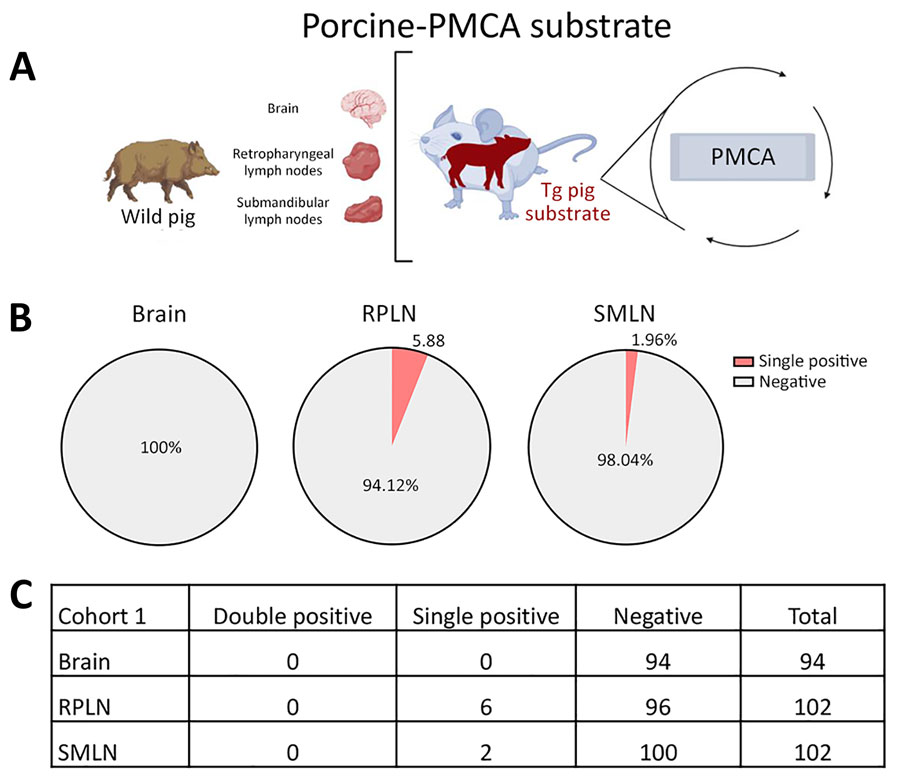
Prion detection using porcine-PMCA in wild pig tissues originating from a CWD-endemic region of Arkansas, USA, in a study of prions in wild pigs (Sus scrofa) from areas with reported chronic wasting disease cases. A) Experimental strategy depicting tissues analyzed in animals from this cohort and PMCA settings. B) Graphs representing percentage of detection per tissue. C) Details on the prion detection data displayed in panel B. PMCA, protein misfolding cyclic amplification; PrP, prion proteins; RPLN, retropharyngeal lymph nodes; SMLN, submandibular lymph nodes; Tg, transgenic.

Figure 2. Prion detection using porcine-PMCA in wild pig tissues originating from a CWD-endemic region of Arkansas, USA, in a study of prions in wild pigs (Sus scrofa) from areas with reported chronic wasting disease cases. A) Experimental strategy depicting tissues analyzed in animals from this cohort and PMCA settings. B) Graphs representing percentage of detection per tissue. C) Details on the prion detection data displayed in panel B. PMCA, protein misfolding cyclic amplification; PrP, prion proteins; RPLN, retropharyngeal lymph nodes; SMLN, submandibular lymph nodes; Tg, transgenic.

Main Article

Page created: December 02, 2024
Page updated: December 22, 2024
Page reviewed: December 22, 2024
The conclusions, findings, and opinions expressed by authors contributing to this journal do not necessarily reflect the official position of the U.S. Department of Health and Human Services, the Public Health Service, the Centers for Disease Control and Prevention, or the authors' affiliated institutions. Use of trade names is for identification only and does not imply endorsement by any of the groups named above.
file_external