Volume 31, Number 1—January 2025
Dispatch
Detection of Prions in Wild Pigs (Sus scrofa) from Areas with Reported Chronic Wasting Disease Cases, United States
Cite This Article
Citation for Media
Abstract
Using a prion amplification assay, we identified prions in tissues from wild pigs (Sus scrofa) living in areas of the United States with variable chronic wasting disease (CWD) epidemiology. Our findings indicate that scavenging swine could play a role in disseminating CWD and could therefore influence its epidemiology, geographic distribution, and interspecies spread.
Chronic wasting disease (CWD) is a prion disease of particular concern because of its uncontrolled contagious spread among various cervid species in North America (https://www.usgs.gov/media/images/distribution-chronic-wasting-disease-north-america-0), its recent discovery in Nordic countries (1), and its increasingly uncertain zoonotic potential (2). CWD is the only animal prion disease affecting captive as well as wild animals. Persistent shedding of prions by CWD-affected animals and resulting environmental contamination is considered a major route of transmission contributing to spread of the disease. Carcasses of CWD-affected animals represent relevant sources of prion infectivity to multiple animal species that can develop disease or act as vectors to spread infection to new locations.
Free-ranging deer are sympatric with multiple animal species, including some that act as predators, scavengers, or both. Experimental transmissions to study the potential for interspecies CWD transmissions have been attempted in raccoons, ferrets, cattle, sheep, and North American rodents (3–7). Potential interspecies CWD transmission has also been addressed using transgenic (Tg) mice expressing prion proteins (PrP) from relevant animal species (8). Although no reports of natural interspecies CWD transmissions have been documented, experimental studies strongly suggest the possibility for interspecies transmission in nature exists (3–7). Inoculation and serial passage studies reveal the potential of CWD prions to adapt to noncervid species, resulting in emergence of novel prion strains with unpredicted features (9–11).
Wild pigs (Sus scrofa), also called feral swine, are an invasive population comprising domestic swine, Eurasian wild boar, and hybrids of the 2 species (12). Wild pig populations have become established in the United States (Appendix Figure 1, panel A), enabled by their high rates of fecundity; omnivorous and opportunistic diet; and widespread, often human-mediated movement (13). Wild pigs scavenge carcasses on the landscape and have an intimate relationship with the soil because of their routine rooting and wallowing behaviors (14). CWD prions have been experimentally transmitted to domestic pigs by intracerebral and oral exposure routes (15), which is relevant because wild pigs coexist with cervids in CWD endemic areas and reportedly prey on fawns and scavenge deer carcasses. Considering the species overlap in many parts of the United States (Appendix Figure 1, panel B), we studied potential interactions between wild pigs and CWD prions.
We screened pig tissues by using the protein misfolding cyclic amplification (PMCA) technique (Appendix). To screen for CWD and porcine-adapted CWD prions in wild pigs, we analyzed the sensitivity and specificity of PMCA to detect prions from different animal species. To evaluate, we first tested the PMCA efficiency of deer CWD prions in both deer and pig substrates. We found the in vitro replication of CWD prions was highly efficient in the deer substrate, and detectable even at high (10−11) dilutions in a first PMCA round (Appendix Figure 2). As a counterpart, we used a porcine-adapted PrPSc (scrapie isoform of infectious prion proteins) pool generated through >10 PMCA rounds in Tg002 substrate (Appendix Figure 3). That inoculum displayed limited PMCA seeding activity after 1 round, confirming the ability of our in vitro prion replication systems to discriminate between homologous (deer) and heterologous (porcine) PrPSc sources (Appendix Figure 2, panel A).
The specificity of our cervid and porcine PMCA systems was further confirmed when using the pig-derived PMCA substrate (Appendix Figure 2, panel B). Specifically, although we detected no amplification in the system when using the CWD seeds, the porcine-adapted PMCA product displayed great amplification. Overall, those results demonstrated that PMCA can be used to identify prions from deer and porcine sources, provide species specificity, and potentially enable tracking of the source of the infectious agent in tissues collected from wild pigs.
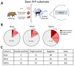
We then investigated the interaction between wild pigs and CWD prions in tissues from animals trapped in areas with different CWD epidemiology. The first cohort of pigs (cohort 1) included animals trapped in Newton and Searcy Counties, Arkansas, USA, where CWD is endemic in free-ranging white-tailed deer (Odocoileus virginianus) and elk (Cervus elaphus canadensis) (https://www.agfc.com/hunting/deer/chronic-wasting-disease/cwd-in-arkansas). The tissues considered for this screening included retropharyngeal lymph nodes (RPLN) and submandibular lymph nodes (SMLN) because those sites have been described to replicate prions shortly after infection in multiple animal species (Appendix). We also tested brainstem samples from the same animals. The first part of the screening involved the use of deer PrP substrate to assess for the potential exposure of wild pigs to CWD prions. We analyzed all samples in duplicate. We were able to detect CWD seeding activity in several wild pig lymphoid tissues and a higher proportion in the SMLN (37.26%) compared with RPLN (16.67%) (Figure 1). As expected, brains displayed a lower detection; just 14.90% of the specimens provided positive signals (Figure 1). We considered a tissue as positive for CWD prions if we noted positive signals in >1 of the 2 replicates. Along that line, a fraction (12.75%) of PMCA-positive SMLN tissues provided positive PMCA results in both replicates, suggesting a higher concentration of CWD prions in SMLN than RPLN or brains (≈3%) (Figure 1). Of note, the CWD infectivity titers in wild pig tissues appeared to be limited because just a fraction of Tg1536 mice (expressing the deer prion protein) inoculated with selected specimens displayed subclinical prion infection (Appendix Table 1, Figure 4).
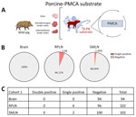
Screening of the same tissues using porcine-PMCA substrate provided a considerably lower number of positive results (Figure 2). Although 5.88% of wild pigs were porcine-PMCA positive at RPLNs, only 1.96% were positive at SMLNs. Of note, none of the tissues that tested PMCA-positive using the porcine substrate provided positive signals in both replicates, suggesting low quantities of seeding-competent porcine prions (Figure 2). None of the brains from cohort 1 provided PMCA seeding activities when the porcine substrate was used, thus confirming those results (Figure 2). Moreover, bioassays in Tg002 mice expressing the porcine prion protein resulted in no transmission (Appendix Table 2).
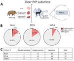
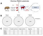
Next, we conducted a screening on a second cohort of animals (cohort 2) collected in Texas, USA. Pigs from cohort 2 were retrieved from counties where either no or low-prevalence CWD had been reported in wild deer. The cervid-adapted PMCA analysis revealed that 15.79% of animals tested positive for CWD prions in RPLNs (Figure 3). Brains (13.16%) and SMLN (8.10%) were also positive, albeit in a lower percentage than for RPLN (Figure 3). Regardless, the overall proportion of PMCA-positive tissues was considerably lower compared with those found for cohort 1, in line with the low prevalence of CWD in free-ranging cervids in the Texas study region. In agreement with the presumably lower exposure of CWD prions for pigs from cohort 2, none of the tissues provided PMCA-positive signals when evaluated in the porcine PMCA system (Figure 4).
In summary, results from this study showed that wild pigs are exposed to cervid prions, although the pigs seem to display some resistance to infection via natural exposure. Future studies should address the susceptibility of this invasive animal species to the multiple prion strains circulating in the environment. Nonetheless, identification of CWD prions in wild pig tissues indicated the potential for pigs to move prions across the landscape, which may, in turn, influence the epidemiology and geographic spread of CWD.
Ms. Soto is a research associate at the Department of Neurology, McGovern Medical School, The University of Texas Health Science Center in Houston, Texas, USA, and is a PhD student at Universidad Bernardo O’Higgins in Santiago, Chile. Her research interests focus on the transmission and spread of chronic wasting disease and the various factors that contribute to it.
Acknowledgments
This project was made possible by the Arkansas Game and Fish Commission, especially Jennifer Ballard, and sample collection was conducted in Arkansas by S. Clarke, C. Smith, W. Wright, and R. Norton. The views and opinions expressed herein are those of the authors and do not necessarily reflect the views or policies of the Arkansas Game and Fish Commission.
This work was supported by United States Department of Agriculture, Animal Plant Health Inspection Service grant nos. 22-7100-0453-CA UTH and AP20WSHQ0000C014, and National Institutes of Health, National Institute of Allergy and Infectious Diseases grant no. 1R01AI132695 to R.M.
R.M. is listed as an inventor in one patent describing the PMCA technique. All other authors have no conflicts to disclose.
Author contributions: P.S. executed most of the experiments described in this article, and prepared the final version of the figures. F.B.-R. and R.B. participated in the screening of feral swine tissues. T.H.S. contributed with western blot analyses of PMCA products. M.J.B., P.W., and C.T. collected wild pig tissues. T.R.S. participated in tissue analyses. J.G. and G.T. provided the original breeder for the transgenic mice used in this study. T.N. provided critical advice on experimental design. V.R.B. and R.M. designed the study and were responsible for funding. R.M. supervised the work and wrote the first draft of the manuscript. All the authors approved the final version of this article.
References
- Benestad SL, Mitchell G, Simmons M, Ytrehus B, Vikøren T. First case of chronic wasting disease in Europe in a Norwegian free-ranging reindeer. Vet Res. 2016;47:88. DOIPubMedGoogle Scholar
- Tranulis MA, Tryland M. The zoonotic potential of chronic wasting disease—a review. Foods. 2023;12:824. DOIPubMedGoogle Scholar
- Moore SJ, Smith JD, Richt JA, Greenlee JJ. Raccoons accumulate PrPSc after intracranial inoculation of the agents of chronic wasting disease or transmissible mink encephalopathy but not atypical scrapie. J Vet Diagn Invest. 2019;31:200–9. DOIPubMedGoogle Scholar
- Sigurdson CJ, Mathiason CK, Perrott MR, Eliason GA, Spraker TR, Glatzel M, et al. Experimental chronic wasting disease (CWD) in the ferret. J Comp Pathol. 2008;138:189–96. DOIPubMedGoogle Scholar
- Greenlee JJ, Nicholson EM, Smith JD, Kunkle RA, Hamir AN. Susceptibility of cattle to the agent of chronic wasting disease from elk after intracranial inoculation. J Vet Diagn Invest. 2012;24:1087–93. DOIPubMedGoogle Scholar
- Cassmann ED, Moore SJ, Greenlee JJ. Experimental oronasal transmission of chronic wasting disease agent from white-tailed deer to Suffolk sheep. Emerg Infect Dis. 2021;27:3156–8. DOIPubMedGoogle Scholar
- Heisey DM, Mickelsen NA, Schneider JR, Johnson CJ, Johnson CJ, Langenberg JA, et al. Chronic wasting disease (CWD) susceptibility of several North American rodents that are sympatric with cervid CWD epidemics. J Virol. 2010;84:210–5. DOIPubMedGoogle Scholar
- Herbst A, Wohlgemuth S, Yang J, Castle AR, Moreno DM, Otero A, et al. Susceptibility of beavers to chronic wasting disease. Biology (Basel). 2022;11:667. DOIPubMedGoogle Scholar
- Morales R, Abid K, Soto C. The prion strain phenomenon: molecular basis and unprecedented features. Biochim Biophys Acta. 2007;1772:681–91. DOIPubMedGoogle Scholar
- Morales R. Prion strains in mammals: Different conformations leading to disease. PLoS Pathog. 2017;13:
e1006323 . DOIPubMedGoogle Scholar - Otero A, Duque Velasquez C, McKenzie D, Aiken J. Emergence of CWD strains. Cell Tissue Res. 2023;392:135–48. DOIPubMedGoogle Scholar
- Smyser TJ, Tabak MA, Slootmaker C, Robeson MS II, Miller RS, Bosse M, et al. Mixed ancestry from wild and domestic lineages contributes to the rapid expansion of invasive feral swine. Mol Ecol. 2020;29:1103–19. DOIPubMedGoogle Scholar
- Bevins SN, Pedersen K, Lutman MW, Gidlewski T, Deliberto TJ. Consequences associated with the recent range expansion of nonnative feral swine. Bioscience. 2014;64:291–9. DOIGoogle Scholar
- Graves HB. Behavior and ecology of wild and feral swine (Sus scrofa). J Anim Sci. 1984;58:482–92. DOIGoogle Scholar
- Moore SJ, West Greenlee MH, Kondru N, Manne S, Smith JD, Kunkle RA, et al. Experimental transmission of the chronic wasting disease agent to swine after oral or intracranial inoculation. J Virol. 2017;91:e00926–17. DOIPubMedGoogle Scholar
Figures
Cite This ArticleOriginal Publication Date: December 17, 2024
Table of Contents – Volume 31, Number 1—January 2025
| EID Search Options |
|---|
|
|
|
|
|
|


