Skip directly to site content Skip directly to page options Skip directly to A-Z link Skip directly to A-Z link Skip directly to A-Z link

Volume 31, Number 10—October 2025

Dispatch

Fatal Pneumocephalus Caused by Hypervirulent Klebsiella pneumoniae, Germany

Nico Gläser1, Nina Schwab1, Yvonne Pfeifer, Anika Wahl, Martin A. Fischer, Anja Osterloh, Thomas F.E. Barth, Armin Imhof, Jochen Breitruck, Andreas Essig, and Jürgen Benjamin HagemannComments to Author 
Author affiliation: Ulm University Hospital, Ulm, Germany (N. Gläser, N. Schwab, A. Osterloh, T.F.E. Barth, A. Imhof, J. Breitruck, A. Essig, J.B. Hagemann); Robert Koch Institute, Wernigerode, Germany (Y. Pfeifer, A. Wahl, M.A. Fischer).

Main Article

Figure 1

Radiologic and pathologic findings from a fatal pneumocephalus case caused by hypervirulent Klebsiella pneumoniae in Germany. A) Computed tomography imaging of the neurocranium revealing gas inclusions both in the arterial and venous system and the inner and outer cerebrospinal fluid spaces consistent with an intravital pneumocephalus. B) Postmortem coronal brain section revealing infiltrates of the leptomeninges and signs of global hypoxic-ischemic brain damage including blurred demarcation between the white and gray matter and discoloration. C) Microscopic appearance of purulent meningitis with bacilli accumulating in the leptomeninges. Hematoxylin and eosin stain. Scale bar indicates 50 µm.

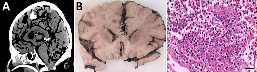
Figure 1. Radiologic and pathologic findings from a fatal pneumocephalus case caused by hypervirulent Klebsiella pneumoniae in Germany. A) Computed tomography imaging of the neurocranium revealing gas inclusions both in the arterial and venous system and the inner and outer cerebrospinal fluid spaces consistent with an intravital pneumocephalus. B) Postmortem coronal brain section revealing infiltrates of the leptomeninges and signs of global hypoxic-ischemic brain damage including blurred demarcation between the white and gray matter and discoloration. C) Microscopic appearance of purulent meningitis with bacilli accumulating in the leptomeninges. Hematoxylin and eosin stain. Scale bar indicates 50 µm.

Main Article

1These first authors contributed equally to this article.

Page created: August 18, 2025
Page updated: September 25, 2025
Page reviewed: September 25, 2025
The conclusions, findings, and opinions expressed by authors contributing to this journal do not necessarily reflect the official position of the U.S. Department of Health and Human Services, the Public Health Service, the Centers for Disease Control and Prevention, or the authors' affiliated institutions. Use of trade names is for identification only and does not imply endorsement by any of the groups named above.
file_external